. 2022 Aug 5;71(3):399-410.
doi: 10.1538/expanim.22-0011. Epub 2022 May 17.

Calcium carbonate supplementation causes motor dysfunction

Affiliations

Calcium carbonate supplementation causes motor dysfunction

Ami Sugiura et al. Exp Anim. .

Abstract

We previously showed that a diet containing calcium carbonate causes impairments in spatial and recognition memory in mice. In this study, we investigated the effects of calcium carbonate supplementation on motor function. Motor function was determined using different tests that have been used to analyze different aspects of Parkinsonism. A catalepsy test for akinesia; a muscular strength assessment, pole test, beam-walking test, and gait analysis for motor coordination and balance assessment; and an open-field test for locomotor activity assessment were performed. The mice were fed diets containing 0.6% or 1.0% calcium carbonate for eight weeks, after which they were evaluated for motor functions. The diets containing calcium carbonate caused significant motor dysfunction, as revealed by the different tests, although the spontaneous locomotor activity did not change. Calcium carbonate supplementation decreased the dopamine content in the basal ganglia, including the striatum and substantia nigra, and the number of tyrosine hydroxylase-positive neurons in the substantia nigra. In addition, administration of L-dopa led to at least a partial recovery of motor dysfunction, suggesting that calcium carbonate supplementation causes motor dysfunction by decreasing the dopamine content in the basal ganglia. These results suggest that mice with calcium carbonate-induced motor dysfunction may be useful as a new animal model for Parkinson's disease and Huntington's disease.

Keywords: Parkinson’s disease; calcium carbonate supplements; dopaminergic neurons; motor dysfunction.


Conflict of interest statement

The authors declare no conflict of interest.

Figures
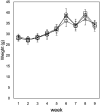
Fig. 1.
Fig. 1.
Influence of calcium carbonate supplementation on body weight. Mice were provided the control diet (○), 0.6% calcium carbonate diet (×), or 1.0% calcium carbonate diet (□). The values of eight mice are presented as means ± SD (n=8).
Fig. 2.
Fig. 2.
Effect of calcium carbonate supplementation on the catalepsy test (a), pole test (b), and beam-walking test findings (c). (a) The duration of catalepsy was measured when the forepaws of mice were placed on a horizontal bar. (b) The time reorient with the head pointed downward in the pole test was measured after the mice were placed with their head oriented upward on the pole. (c) The number of times the mice slipped off the beam was measured when they were made to walk across a narrow beam. The values of eight mice are presented as means ± SD (n=8). Asterisks indicate statistically significant differences relative to the control (P<0.05).
Fig. 3.
Fig. 3.
Effect of calcium carbonate supplementation on the gait analysis (a), locomotor activity (b), and buried pellet test (c) findings. (a) In the gait analysis, paint was applied to the forepaws (red) and hind paws (blue) of the mice, and the mice were then allowed to walk on a piece of white paper to record the placement pattern of their footprints. The step length, step width of the hind paw and forepaw, and overlap asymmetry were evaluated (Table 3). (b) The number of line crosses of the mice within 5 min was measured. (c) After the mice were placed in the test box where the pellet was buried, the latency to pellet detection was measured. The values of eight mice are presented as means ± SD (n=8). Asterisks indicate statistically significant differences relative to the control (P<0.05).
Fig. 4.
Fig. 4.
Tyrosine hydroxylase (TH) expression in the striatum of the mice fed the control and calcium carbonate diets. (a) Western blotting of the basal ganglia extracted from the mice fed the control diet (1), 0.6% calcium carbonate diet (2), or 1.0% calcium carbonate diet (3). (b) Immunostaining of TH in the substantial nigra (upper and middle panels) and the striatum (lower panel) of the mice fed the control diet, 0.6% calcium carbonate diet, or 1.0% calcium carbonate diet. TH-positive neurons are indicated by an arrowhead. Scale bar=20 (middle panel) and 100 µm (lower and upper panels). (c) The number of TH-positive neurons in the substantial nigra was calculated in five different fields for each mouse. (d) The neuronal cell morphology in the entire (upper panel) and CA1 regions (lower panel) in the hippocampus of the mice fed with the control diet and 1.0% calcium carbonate diet was observed with hematoxylin-eosin staining. Scale bar=17.5 and 175 µm.
Fig. 5.
Fig. 5.
Monoamine content in the striatum of the mice fed the control and calcium carbonate diets. The contents of noradrenaline (a), serotonin (b), L-dopa (c), and dopamine (d) in the striatum were measured as described in the Materials and Methods. The values of five mice are presented as means ± SD (n=5). Asterisks indicate statistically significant differences relative to the control (P<0.05).
Fig. 6.
Fig. 6.
Changes in the expression of dopamine-related proteins (a) and calcium-binding proteins (b) in the basal ganglia of the mice fed the control and calcium carbonate diets. The mRNA expression levels were measured using real-time polymerase chain reaction. (c) Oxidative stress in the brain of the mice fed the control and calcium carbonate diets was estimated on the basis of the malondialdehyde content and the expression levels of antioxidant enzymes. The values of five to eight mice are presented as means ± SD. Asterisks indicate statistically significant differences relative to the control (P<0.05).
Fig. 7.
Fig. 7.
Effect of L-dopa administration on calcium carbonate-induced motor dysfunction. The mice fed the control, 0.6% calcium carbonate, and 1.0% calcium carbonate diets for 2 months were administered L-dopa, and the catalepsy (a) and pole (b) tests were performed. Open and closed bars show the values of the treated and untreated mice, respectively. The values of five to eight mice are presented as means ± SD. Asterisks indicate statistically significant differences relative to the control (P<0.05).

Similar articles

Cited by

References

    1. Popugaeva E, Pchitskaya E, Bezprozvanny I. Dysregulation of intracellular calcium signaling in Alzheimer’s disease. Antioxid Redox Signal. 2018; 29: 1176–1188. doi: 10.1089/ars.2018.7506 - DOI - PMC - PubMed
    1. Neher E, Sakaba T. Multiple roles of calcium ions in the regulation of neurotransmitter release. Neuron. 2008; 59: 861–872. doi: 10.1016/j.neuron.2008.08.019 - DOI - PubMed
    1. Zucker RS. Calcium- and activity-dependent synaptic plasticity. Curr Opin Neurobiol. 1999; 9: 305–313. doi: 10.1016/S0959-4388(99)80045-2 - DOI - PubMed
    1. Berridge MJ. Neuronal calcium signaling. Neuron. 1998; 21: 13–26. doi: 10.1016/S0896-6273(00)80510-3 - DOI - PubMed
    1. Szydlowska K, Tymianski M. Calcium, ischemia and excitotoxicity. Cell Calcium. 2010; 47: 122–129. doi: 10.1016/j.ceca.2010.01.003 - DOI - PubMed