Review
. 2022 Oct;47(10):875-891.
doi: 10.1016/j.tibs.2022.04.011. Epub 2022 May 16.

Looking lively: emerging principles of pseudokinase signaling

Affiliations
Review

Looking lively: emerging principles of pseudokinase signaling

Joshua B Sheetz et al. Trends Biochem Sci. 2022 Oct.

Abstract

Progress towards understanding catalytically 'dead' protein kinases - pseudokinases - in biology and disease has hastened over the past decade. An especially lively area for structural biology, pseudokinases appear to be strikingly similar to their kinase relatives, despite lacking key catalytic residues. Distinct active- and inactive-like conformation states, which are crucial for regulating bona fide protein kinases, are conserved in pseudokinases and appear to be essential for function. We discuss recent structural data on conformational transitions and nucleotide binding by pseudokinases, from which some common principles emerge. In both pseudokinases and bona fide kinases, a conformational toggle appears to control the ability to interact with signaling effectors. We also discuss how biasing this conformational toggle may provide opportunities to target pseudokinases pharmacologically in disease.

Keywords: allostery; cell signaling; conformational disruptor; kinase; protein conformation; pseudokinase.


Conflict of interest statement

Declaration of interests The authors declare no conflicts of interest.

Figures

Figure 1
Figure 1. Kinases and conformational toggling
(A) Key residues and sequence motifs in a protein kinase domain. An abbreviated primary sequence of the bona fide kinase protein kinase A (PKA) is shown, with secondary structural elements represented as black arrows (β strands) or grey rectangles (α helices), and marked. The key motifs are indicated and boxed in red. The activation loop is marked as a thick green line and catalytic loop as thick black line. (B,C) Conformational transition between active (B) and inactive (C) conformations of IRK (PDB IDs: 1IR3 [101] and 1IRK [102]). Kinase domains are colored slate blue, and the N-lobe colored according to the backbone position root mean square deviation (RMSD) of alpha carbon positions between active and inactive conformation states – red representing the greatest changes. Activation loops are green, with YxxxYY motif tyrosine side-chains shown, phosphorylated in (B). The β3 lysine (K) and αC glutamate (E) are marked, with the interaction shown by a dotted line in (B). Bound AMP-PNP and Mg2+ (brown) are shown in (B), but are absent in (C). The αC position is assigned ‘in’ (active) or ‘out’ (inactive). (D,E) Transition between the active (D) and inactive (E) EGFR kinase domain conformations (PDB IDs: 3VJO [103] and 2GS7 [104]), labeled as in (B) and (C). Both structures have AMP-PNP bound. The Mg2+ ion in (D) is positioned based on its location in (E). (F) Activation of the EGFR kinase domain via the purple ‘αC patch’. The (active) receiver molecule within the asymmetric dimer is the same as in (D), and the activator is yellow. Interfacial residues in the receiver N-lobe are shown (purple), and constitute the ‘αC patch’. From PDB ID 3VJO [103]. (G) Activation of CDK2 by cyclin A, represented as for EGFR in (F), with the αC patch highlighted (PDB ID: 1FIN [44]). (H,I) Conformational toggling of the Aurora A (AurA) kinase domain from the active ATP-bound conformation (H) to an inactive conformation (I) when bound to the conformational disruptor CD532. Labels are as in (B). PDB IDs are 1MQ4 [105] and 4J8M [76] respectively. AurA binds N-MYC in the active conformation (H), but not the disrupted conformation (I).
Figure 2
Figure 2. Conformational toggling of pseudokinases
(A,B) Conformational transition between active-like (A) and inactive-like (B) conformations of MLKL (PDB IDs: 7JW7 [47] and 7JXU [47]), labeled and colored as in Figure 1, with N-lobe colored according to the RMSD between states in an N-lobe overlay. (C) Allosteric activation of the bona fide protein kinase MST4 by MO25α (PDB ID: 4FZA [48]), resembling EGFR and CDK2 activation in Figure 1. MO25α (yellow) binds to residues in the MST4 N-lobe that constitute the αC patch (C), which in turn coincides with the most variable region in the MST4 N-lobe between active and inactive conformations. (D) MST4 kinase domain in an inactive conformation (PDB ID: 3GGF [106]), bound to a quinazoline inhibitor [106], which is shown in orange. (E) Conformational modulation of the pseudokinase STRADα by MO25α to activate LKB1. MO25α binds the αC patch of STRADα to stabilize it in the active-like ATP-bound conformation. The two molecules form a heterotrimer with the bona fide kinase LKB1/STK11 (pink) with STRADα recognizing LKB1 like a pseudosubstrate and allosterically activating it. PDB ID: 2WTK [35]. (F,G) Conformational transition between active-like (F) and inactive-like (G) TRIB1 (PDB IDs: 5CEM [52] and 6DC0 [51]), as shown for MLKL in (A). In the active-like state (F), TRIB1 binds the degron of C/EBP family transcription factors (shown in sand color) like a pseudosubstrate, and promotes release of the C-terminal fragment of TRIB1 (orange in G) that docks adjacent to αC – near the αC patch – in the inactive-like conformation.
Figure 3
Figure 3. Conformational switching in RAS: an analogy for pseudokinases?
(A,B) RAS as a prototypical G protein conformational switch. RAS is represented in cartoon form, colored according to backbone position RMSD when GTP- and GDP-bound forms are overlaid (blue is no difference, red is maximum difference: ~5Å). Regions undergoing the greatest conformational changes were originally named switch I and switch II [107] – as labelled. Bound nucleotides are shown in black sticks and marked, and Mg2+ ions are brown spheres. Active RAS is from PDB ID 4G0N [108] and inactive RAS from PDB ID 4Q21 [107]. (C) Active GTP-bound RAS binds effector proteins phospholipase Cε and RAF (through its RAS-binding domain or RBD) via switch I. Interfacial residues in RAS are colored purple, and correspond with switch I. PLCε is shown as orange ribbons (PBD ID: 2C5L [109]) and RAF-RBD as green ribbons (PDB ID: 4G0N [108]). (D) Active GTP-bound RAS uses both switch I and switch II to bind phosphatidylinositol-3-kinase (PI3K) and Nore1A, with interfacial residues (purple) coinciding with the two switch regions just as the kinase αC patch coincides with regions of maximum change in the kinase N-lobe. PI3K is shown in pale yellow ribbons (PDB ID: 1HE8 [110]) and Nore1A is shown as pale blue ribbons (PDB ID: 3DDC [111]).
Figure 4
Figure 4. Resilience of pseudokinase ATP binding
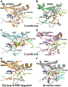
ATP binding sites are shown for a series of pseudokinase domains that retain ATP binding (A-E) and for PKA (F) for comparison. Bound ATP, AMP-PNP or ATPγS is shown. The part of the activation loop shown is colored green (as is the DFG Asp when present), and preserved ATP-interacting side-chains are shown in black sticks. ‘Compensatory’ side-chains that make up for lost ATP-interacting residues by contacting the nucleotide as described in the text are colored orange. Mg2+ is shown as brown sphere in (B, C, F), but is absent in (A, D, E), which show cation-independent ATP binding.
Figure 5
Figure 5. Modulating pseudokinases with small molecules
(A-C) Comparison of the PKA ATP-binding site (A) with those of the ROR2 (B) and ROR1 (C), shown in the same orientation. Hydrophobic side-chains occluding the ROR2 and ROR1 ATP-binding sites are shown as sticks and transparent spheres. The activation loop section containing the YxxxYY motif that also contributes to active site occlusion is green. Ponatinib (red) binds ROR1 (but not ROR2) [63] in a cavity above the occluded (vestigial) ATP-binding site (C: PDB ID: 6TU9 [63]). Similar cavities exist in ROR2 (B: PDB ID: 3ZZW), RYK, and PTK7, that may also be targetable. ATP-binding motifs marked in Figure 1A are shown in each binding site (β3 lysine, αC glutamate, HRD and DFG aspartates). (D) Conformational disruption of KSR2 by APS-2-79 (orange sticks/spheres). The KSR2 pseudokinase domain is blue, with APS-2-79-induced conformational changes colored red according to RMSD changes between drug- and ATP-bound states (comparing PDB IDs: 5KKR [77] and 2Y4I [19]). KSR2 forms an inhibitory complex with MEK (grey surface representation) that is stabilized by the APS-2-79-induced ‘lock’ in KSR2 (residues I809-Q814: colored red), ordered by compound binding [77], APS-2-79-induced alterations on the opposite face of KSR2 appear to inhibit its association with RAF (transparent green surface, modeled based on the B-RAF dimer in PDB ID: 1UWH [112]). (E) N-lobe-mediated contacts between the JH2 pseudokinase domain of TYK2 (magenta) and the JH1 (bona fide) kinase domain in the same TYK2 molecule (sand), from PDB ID: 4OLI [82]. The activation loop is colored green and each ATP-binding site is occupied by an ATP-competitive TYK2 inhibitor that binds both domains [82]. Selective binding of deucravacitinib/BMS-986165 to the JH2 domain [80] stabilizes this configuration and indirectly inhibits JH1 activation as described in the text.
Figure I.
Figure I.. Old Domain, New Tricks.
(A) PKA represents a prototypical protein kinase (PDB ID: 1ATP [113]). The kinase domain is shown as a grey cartoon. Bound ATP and peptide substrates are shown as black and cyan sticks respectively, and Mg2+ ions are shown as brown spheres. (B) POMK transfers the γ-phosphate from ATP to the mannose unit of glycan-modified α-dystroglycan. The kinase domain is shown as a pale green cartoon. A trisaccharide substrate, colored cyan, traverses the canonical peptide binding site to place the mannose O6 hydroxyl in a phosphoacceptor position next to ATP (present as ADP in the crystal) and the catalytic base aspartate (PDB ID: 5GZ9 [114]). (C) SelO transfers AMP from ATP onto Ser/Thr/Tyr residues of redox regulatory proteins. The ATP analog AMP-PNP is present in a ‘flipped’ orientation compared to the canonical binding mode in active protein kinases – also seen in the secreted Fam20A pseudokinase [72]. The core kinase domain is colored pale blue, and the SelO-specific sequences are light gray (PDB ID: 6EAC [24]). (D) The NiRAN domain from nsp12 in the SARS-CoV-2 replication-transcription complex is shown (PDB ID: 6XEZ [92]). The NiRAN domain resembles SelO structurally and in its ‘flipped’ mode of nucleotide binding, with Mg2+-ADP bound in this structure. (E) The Legionella effector protein SidJ polyglutamylates SidE family ubiquitin ligases during viral amplification. The core pseudokinase domain is shown as a pale pink cartoon, and the SidJ N- and C-terminal domains (NTD and CTD respectively) are colored light yellow (NTD) and gray (CTD). The bound yeast calmodulin (CaM), which activates SidJ, is colored light cyan (PDB ID: 7MIR [23]).

Similar articles

Cited by

References

    1. Burnett G and Kennedy EP (1954) The enzymatic phosphorylation of proteins. J. Biol. Chem 211, 969–980 - PubMed
    1. Pawson T and Scott JD (2005) Protein phosphorylation in signaling--50 years and counting. Trends Biochem. Sci 30, 286–290 - PubMed
    1. Adams JA (2001) Kinetic and catalytic mechanisms of protein kinases. Chem. Rev 101, 2271–2290 - PubMed
    1. Manning G, et al. (2002) The protein kinase complement of the human genome. Science 298, 1912–1934 - PubMed
    1. Kwon A, et al. (2019) Tracing the origin and evolution of pseudokinases across the tree of life. Sci. Signal 12, eaav3810. - PMC - PubMed

Publication types

MeSH terms

Substances

LinkOut - more resources