

Comparison of long-term visual and anatomical outcomes between internal limiting membrane flap and peeling techniques for macular holes with a propensity score analysis
- PMID: 35585135
- PMCID: PMC10102159
- DOI: 10.1038/s41433-022-02103-5
Comparison of long-term visual and anatomical outcomes between internal limiting membrane flap and peeling techniques for macular holes with a propensity score analysis
Abstract
Objectives: To compare visual and anatomical outcomes between internal limiting membrane (ILM) flap (IF) and peeling (IP) techniques for full-thickness macular holes (FTMHs).
Methods: A retrospective case series with propensity-score matching (PSM). Patients with a minimum 12 months follow-up were divided into IF and IP groups and matched based on FTMH size and preoperative best-corrected visual acuity (BCVA). BCVA and optical coherence tomography (OCT) findings were obtained to assess outer retinal layer integrity, foveal thickness, and foveal displacement.
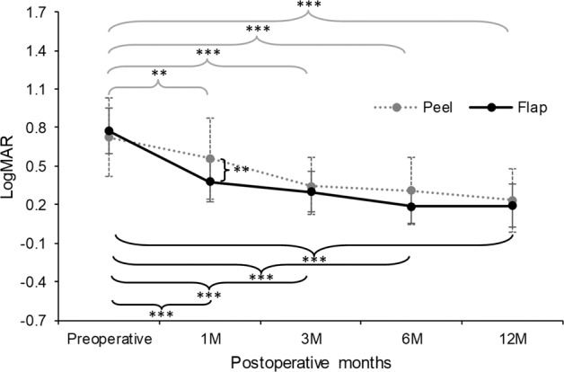
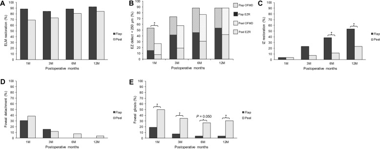
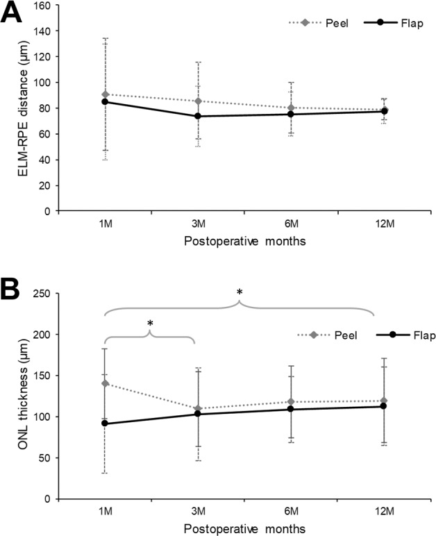
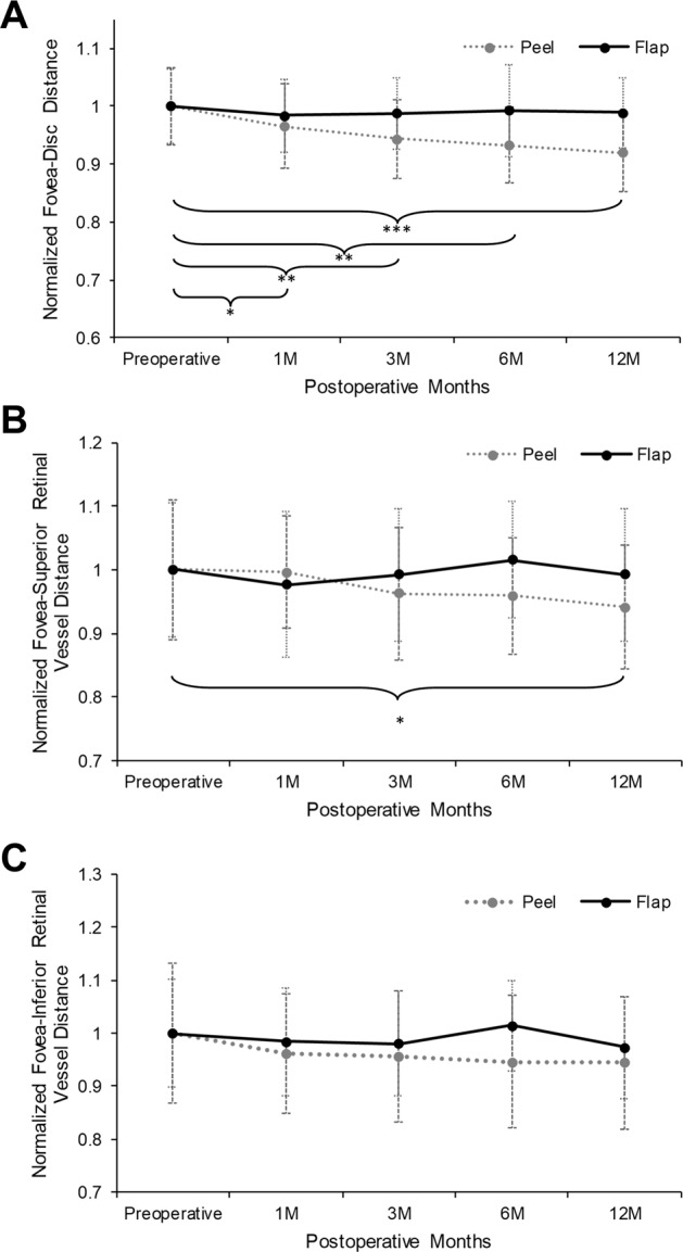
Results: Twenty-six eyes were included in each group after PSM. The IF group showed significantly greater BCVA after 1 month, its corresponding change from preoperative BCVA, proportions of eyes with ellipsoid zone defects <250 μm after 1 month, and interdigitation zone restoration after 6 and 12 months (P = 0.007, 0.038, 0.048, 0.025, and 0.023, respectively), as well as less foveal gliosis after 1, 3, 6, and 12 months (P = 0.020, 0.017, 0.050, and 0.024, respectively). In the IP group, the mean outer nuclear layer thickness significantly decreased at 3 (P = 0.019) and 12 months (P = 0.016) compared to 1 month, and the foveal displacement toward the optic disc was significant after 1, 3, 6, and 12 months (P = 0.049, 0.006, 0.001, and <0.001, respectively).
Conclusions: Compared to IP, IF promoted faster recovery of BCVA and outer retinal layers and was more protective against postoperative foveal thinning and displacement; hence, it should be considered for small and large FTMHs.
© 2022. The Author(s), under exclusive licence to The Royal College of Ophthalmologists.
Conflict of interest statement
The authors declare no competing interests.
Figures
Similar articles
-
Inverted internal limiting membrane flap technique versus complete internal limiting membrane peeling in large macular hole surgery: a comparative study.BMC Ophthalmol. 2020 Jan 6;20(1):11. doi: 10.1186/s12886-019-1294-8. BMC Ophthalmol. 2020. PMID: 31907015 Free PMC article.
-
Comparative study of conventional internal limiting membrane peeling versus temporal inverted internal limiting membrane flap for large macular hole treatment.Indian J Ophthalmol. 2023 Jan;71(1):188-194. doi: 10.4103/ijo.IJO_685_22. Indian J Ophthalmol. 2023. PMID: 36588234 Free PMC article.
-
[Vitrectomy and iOCT-assisted inverted ILM flap technique in patients with full thickness macular holes].Ophthalmologe. 2019 Jul;116(7):617-624. doi: 10.1007/s00347-018-0769-y. Ophthalmologe. 2019. PMID: 30105564 German.
-
Comparative efficacy evaluation of inverted internal limiting membrane flap technique and internal limiting membrane peeling in large macular holes: a systematic review and meta-analysis.BMC Ophthalmol. 2020 Jan 8;20(1):14. doi: 10.1186/s12886-019-1271-2. BMC Ophthalmol. 2020. PMID: 31914954 Free PMC article.
-
Comparison of Different Internal Limiting Membrane Peeling Sizes for Idiopathic Macular Holes: A Systematic Review and Meta-Analysis.Ophthalmic Res. 2023;66(1):1071-1084. doi: 10.1159/000531510. Epub 2023 Aug 16. Ophthalmic Res. 2023. PMID: 37586342 Free PMC article.
Cited by
-
Glial-Plug Proliferation after Inverted Internal Limiting Membrane Flap Technique for Idiopathic Macular Hole.J Ophthalmol. 2022 Sep 30;2022:2919358. doi: 10.1155/2022/2919358. eCollection 2022. J Ophthalmol. 2022. PMID: 36237556 Free PMC article.
-
Retinal displacement after surgery for idiopathic macular hole.Int J Ophthalmol. 2024 Aug 18;17(8):1545-1556. doi: 10.18240/ijo.2024.08.21. eCollection 2024. Int J Ophthalmol. 2024. PMID: 39156782 Free PMC article. Review.
-
Comparison of stereopsis and foveal microstructure after internal limiting membrane peeling and inverted internal limiting membrane flap techniques in patients with macular hole.PLoS One. 2024 Feb 9;19(2):e0297134. doi: 10.1371/journal.pone.0297134. eCollection 2024. PLoS One. 2024. PMID: 38335184 Free PMC article.
-
Comparison of the use of internal limiting membrane flaps versus conventional ILM peeling on post-operative anatomical and visual outcomes in large macular holes.Eye (Lond). 2024 Jul;38(10):1876-1881. doi: 10.1038/s41433-024-03024-1. Epub 2024 Mar 16. Eye (Lond). 2024. PMID: 38493269 Free PMC article.
References
MeSH terms
Grants and funding
LinkOut - more resources
Full Text Sources
Miscellaneous
