. 2022 May 18;22(1):184.
doi: 10.1186/s12905-022-01765-3.

Diagnostic gene biomarkers for predicting immune infiltration in endometriosis

Affiliations

Diagnostic gene biomarkers for predicting immune infiltration in endometriosis

Chengmao Xie et al. BMC Womens Health. .

Abstract

Objective: To determine the potential diagnostic markers and extent of immune cell infiltration in endometriosis (EMS).

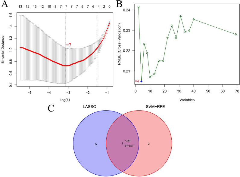
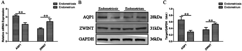
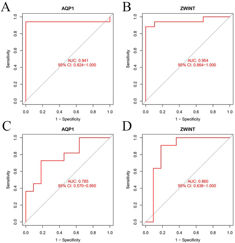
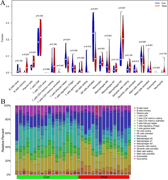
Methods: Two published profiles (GSE7305 and GSE25628 datasets) were downloaded, and the candidate biomarkers were identified by support vector machine recursive feature elimination analysis and a Lasso regression model. The diagnostic value and expression levels of biomarkers in EMS were verified by quantitative reverse transcription polymerase chain reaction (qRT-PCR) and western blotting, then further validated in the GSE5108 dataset. CIBERSORT was used to estimate the composition pattern of immune cell components in EMS.

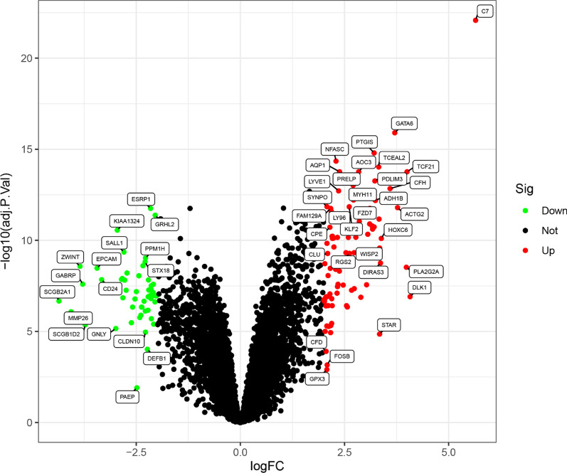
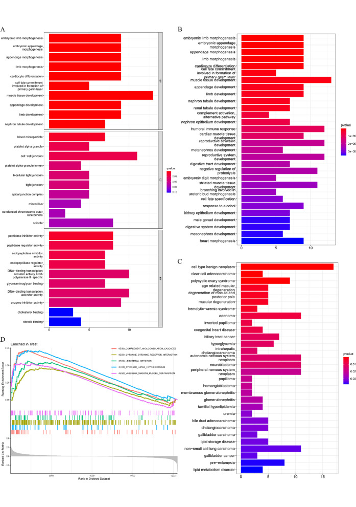
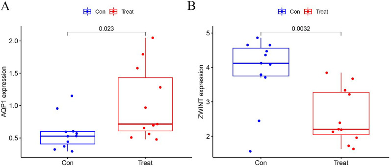
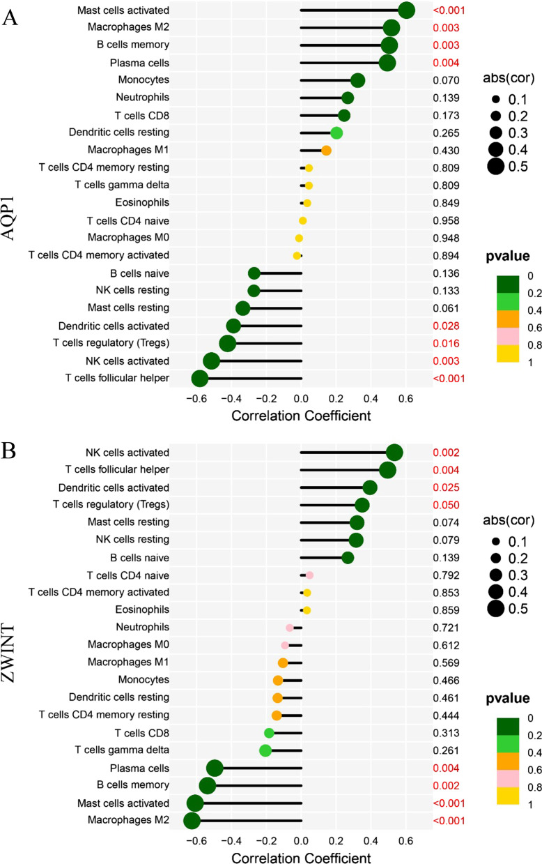
Results: One hundred and fifty-three differential expression genes (DEGs) were identified between EMS and endometrial with 83 upregulated and 51 downregulated genes. Gene sets related to arachidonic acid metabolism, cytokine-cytokine receptor interactions, complement and coagulation cascades, chemokine signaling pathways, and systemic lupus erythematosus were differentially activated in EMS compared with endometrial samples. Aquaporin 1 (AQP1) and ZW10 binding protein (ZWINT) were identified as diagnostic markers of EMS, which were verified using qRT-PCR and western blotting and validated in the GSE5108 dataset. Immune cell infiltrate analysis showed that AQP1 and ZWINT were correlated with M2 macrophages, NK cells, activated dendritic cells, T follicular helper cells, regulatory T cells, memory B cells, activated mast cells, and plasma cells.

Conclusion: AQP1 and ZWINT could be regarded as diagnostic markers of EMS and may provide a new direction for the study of EMS pathogenesis in the future.

Keywords: Biomarker; CIBERSORT; Endometriosis; GEO; Immune infiltration.


Conflict of interest statement

The authors declare that they have no competing interests.

Figures

Fig. 1
Fig. 1
The volcano map of DEGs between EMS and endometrial samples. Each dot represents a gene, and the blue and red dots represent significantly differentially expressed genes. The red dot indicates that the gene expression is up-regulated and the blue dot indicates that the gene expression is down regulated, and the gray dot indicates that there is no significant difference between these genes in EMS
Fig. 2
Fig. 2
GO, KEGG, DO, and GSEA pathway enrichment. A GO enrichment analysis. B KEGG enrichment analysis. C DO enrichment analysis. D GSEA enrichment analysis
Fig. 3
Fig. 3
The process of screening diagnostic biomarker candidates for endometriosis. A Results of the LASSO regression algorithm of the DEGs for EMS. B Results of the SVM-RFE algorithm among the DEGs. C Venn diagram of the two different algorithms
Fig. 4
Fig. 4
Expression of the two biomarkers in the GSE5108 dataset. A AQP1. B ZWINT
Fig. 5
Fig. 5
Results of differential gene expression of AQP1 and ZWINT at the mRNA and protein level between EMS and control samples. A. RT-PCR results; B. Western blotting results; C. Densitometry of the western blot; **P < 0.01
Fig. 6
Fig. 6
The diagnostic effectiveness of the two markers was represented by the receiver operating characteristic (ROC) curve. A AQP1. B ZWINT. C AQP1 in the GSE5108 dataset. D ZWINT in the GSE5108 dataset
Fig. 7
Fig. 7
Visualization and distribution of immune cell infiltrates. A Composition of immune cells in the EMS and control groups. Blue and red colors represent control and EMS samples. B Correlation matrix results of the immune cells
Fig. 8
Fig. 8
Correlation between the two biomarkers and infiltrating immune cells in endometriosis. A AQP1. B ZWINT

Similar articles

Cited by

References

    1. Burney RO, Giudice LC. Pathogenesis and pathophysiology of endometriosis. Fertil Steril. 2012;98(3):511–519. doi: 10.1016/j.fertnstert.2012.06.029. - DOI - PMC - PubMed
    1. Johnson NP, Hummelshoj L, Adamson GD, Keckstein J, Taylor HS, Abrao MS, Bush D, Kiesel L, Tamimi R, Sharpe-Timms KL, et al. World endometriosis society consensus on the classification of endometriosis. Hum Reprod. 2015;32(2):315–324. doi: 10.1093/humrep/dew293. - DOI - PubMed
    1. Rogers PA, Adamson GD, Al-Jefout M, Becker CM, D’ Hooghe TM, Dunselman GA, Fazleabas A, Giudice LC, Horne AW, et al. Research priorities for endometriosis. Reprod Sci. 2015;24(2):202–226. doi: 10.1177/1933719116654991. - DOI - PMC - PubMed
    1. Bonocher CM, Montenegro ML, Rosa E, Silva JC, Ferriani RA, Meola J. Endometriosis and physical exercises: a systematic review. Reprod Biol Endocrinol. 2014;12:4. doi: 10.1186/1477-7827-12-4. - DOI - PMC - PubMed
    1. Dunselman GA, Vermeulen N, Becker C, Calhaz-Jorge C, D’ Hooghe T, De Bie B, Heikinheimo O, Horne AW, Kiesel L, et al. ESHRE guideline: management of women with endometriosis. Hum Reprod. 2014;29(3):400–412. doi: 10.1093/humrep/det457. - DOI - PubMed