

No evidence of bovine leukemia virus proviral DNA and antibodies in human specimens from Japan
- PMID: 35585539
- PMCID: PMC9116711
- DOI: 10.1186/s12977-022-00592-6
No evidence of bovine leukemia virus proviral DNA and antibodies in human specimens from Japan
Abstract
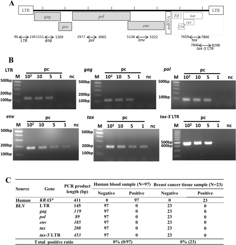
Background: The potential risk and association of bovine leukemia virus (BLV) with human remains controversial as it has been reported to be both positive and negative in human breast cancer and blood samples. Therefore, establishing the presence of BLV in comprehensive human clinical samples in different geographical locations is essential.
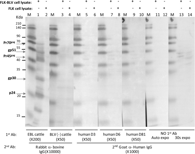
Result: In this study, we examined the presence of BLV proviral DNA in human blood and breast cancer tissue specimens from Japan. PCR analysis of BLV provirus in 97 Japanese human blood samples and 23 breast cancer tissues showed negative result for all samples tested using long-fragment PCR and highly-sensitive short-fragment PCR amplification. No IgG and IgM antibodies were detected in any of the 97 human serum samples using BLV gp51 and p24 indirect ELISA test. Western blot analysis also showed negative result for IgG and IgM antibodies in all tested human serum samples.
Conclusion: Our results indicate that Japanese human specimens including 97 human blood, 23 breast cancer tissues, and 97 serum samples were negative for BLV.
Keywords: Antibody detection; Bovine leukemia virus (BLV); Japanese breast cancers; Japanese human blood; Japanese human sera; PCR.
© 2022. The Author(s).
Conflict of interest statement
The authors declare that they have no competing interests.
Figures

Similar articles
-
Bovine leukaemia virus: rapid detection of proviral DNA by nested PCR in blood and organs of experimentally infected calves.Vet Microbiol. 1994 Nov;42(2-3):191-204. doi: 10.1016/0378-1135(94)90018-3. Vet Microbiol. 1994. PMID: 7886932
-
Proviral detection and serology in bovine leukemia virus-exposed normal cattle and cattle with lymphoma.Can J Vet Res. 1992 Oct;56(4):339-48. Can J Vet Res. 1992. PMID: 1335834 Free PMC article.
-
Risk factors associated with increased bovine leukemia virus proviral load in infected cattle in Japan from 2012 to 2014.Virus Res. 2015 Dec 2;210:283-90. doi: 10.1016/j.virusres.2015.08.020. Epub 2015 Aug 29. Virus Res. 2015. PMID: 26321160
-
Bovine leukemia virus discovered in human blood.BMC Infect Dis. 2019 Apr 2;19(1):297. doi: 10.1186/s12879-019-3891-9. BMC Infect Dis. 2019. PMID: 30940091 Free PMC article.
-
Investigation of the bovine leukemia virus proviral DNA in human leukemias and lung cancers in Korea.J Korean Med Sci. 2005 Aug;20(4):603-6. doi: 10.3346/jkms.2005.20.4.603. J Korean Med Sci. 2005. PMID: 16100451 Free PMC article.
Cited by
-
The Role of Oncogenic Viruses in the Pathogenesis of Sporadic Breast Cancer: A Comprehensive Review of the Current Literature.Pathogens. 2024 May 25;13(6):451. doi: 10.3390/pathogens13060451. Pathogens. 2024. PMID: 38921749 Free PMC article. Review.
-
Bovine Leukemia Virus and Human Breast Cancer: A Review of Clinical and Molecular Evidence.Viruses. 2025 Feb 26;17(3):324. doi: 10.3390/v17030324. Viruses. 2025. PMID: 40143252 Free PMC article. Review.
-
A safe and effective vaccine against bovine leukemia virus.Front Immunol. 2022 Aug 10;13:980514. doi: 10.3389/fimmu.2022.980514. eCollection 2022. Front Immunol. 2022. PMID: 36032174 Free PMC article.
-
The association between infectious agents and breast cancer: a review of the epidemiologic evidence.Breast Cancer Res Treat. 2024 Sep;207(2):235-252. doi: 10.1007/s10549-024-07388-6. Epub 2024 Jul 6. Breast Cancer Res Treat. 2024. PMID: 38971906 Review.
-
Milk Transmission of Mammalian Retroviruses.Microorganisms. 2023 Jul 8;11(7):1777. doi: 10.3390/microorganisms11071777. Microorganisms. 2023. PMID: 37512949 Free PMC article. Review.
References
Publication types
MeSH terms
Substances
LinkOut - more resources
Full Text Sources
