. 2022 May 18;23(1):127.
doi: 10.1186/s12931-022-02045-2.

MAIT cell counts are associated with the risk of hospitalization in COPD

Affiliations

MAIT cell counts are associated with the risk of hospitalization in COPD

Terezia Pincikova et al. Respir Res. .

Abstract

Background: Chronic obstructive pulmonary disease (COPD) is characterized by persistent airflow limitation associated with chronic inflammation in the airways. Mucosal-associated invariant T (MAIT) cells are unconventional, innate-like T cells highly abundant in mucosal tissues including the lung. We hypothesized that the characteristics of MAIT cells in circulation may be prospectively associated with COPD morbidity.

Methods: COPD subjects (n = 61) from the Tools for Identifying Exacerbations (TIE) study were recruited when in stable condition. At study entry, forced expiratory volume in 1 s (FEV1) was measured and peripheral blood mononuclear cells were cryopreserved for later analysis by flow cytometry. Patients were followed for 3 years to record clinically meaningful outcomes.

Results: Patients who required hospitalization at one or more occasions during the 3-year follow-up (n = 21) had lower MAIT cell counts in peripheral blood at study inclusion, compared with patients who did not get hospitalized (p = 0.036). In contrast, hospitalized and never hospitalized patients did not differ in CD8 or CD4 T cell counts (p = 0.482 and p = 0.221, respectively). Moreover, MAIT cells in hospitalized subjects showed a more activated phenotype with higher CD38 expression (p = 0.014), and there was a trend towards higher LAG-3 expression (p = 0.052). Conventional CD4 and CD8 T cells were similar between the groups. Next we performed multi-variable logistic regression analysis with hospitalizations as dependent variable, and FEV1, GOLD 2017 group, and quantity or activation of MAIT and conventional T cells as independent variables. MAIT cell count, CD38 expression on MAIT cells, and LAG-3 expression on both MAIT and CD8 T cells were all independently associated with the risk of hospitalization.

Conclusions: These findings suggest that MAIT cells might reflect a novel, FEV1-independent immunological dimension in the complexity of COPD. The potential implication of MAIT cells in COPD pathogenesis and MAIT cells' prognostic potential deserve further investigation.

Keywords: COPD; Human; Immune activation; MAIT cells; T cells.


Conflict of interest statement

The authors declare that they have no competing interests.

Figures
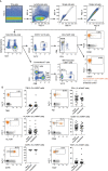
Fig. 1
Fig. 1
Gating strategy. A Gating strategy to identify peripheral blood CD4 T cells, classical CD8 T cells, MAIT cells and expression of the activation markers CD38 and LAG-3 on MAIT cells. Gates for CD38 and LAG-3 expression on MAIT cells (orange) were set based on the expression on total T cells (in grey) when a clear negative and positive population could be identified or based on the unstained control (black) otherwise. MAIT cells were identified as CD4-Vα7.2 + CD161 + T cells. For quantification of classical CD8 T cells, MAIT cells were gated out. B Phenotypic comparison of MAIT cells in COPD patients requiring hospitalization or not during the 3-year follow-up. Representative flow cytometry staining of TIM-3, PD1, HLA-DR, CD56, CCR5 and TIGIT on MAIT cells (orange) using the unstained control (black) or the total T cells (grey) as controls to set the gate. The scatter plots display the expression of each marker (median ± IQR) on MAIT cells between Hospitalized (n = 21) and Never Hospitalized (n = 40) COPD patients during the 3-year follow-up. The Mann–Whitney U Test was used to evaluate statistical differences between groups
Fig. 2
Fig. 2
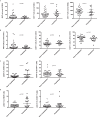
MAIT cell count and expression of the activation marker CD38 are associated with the risk of hospitalization. MAIT cell count, CD8 T cell count, CD4 T cell count (A), MAIT cell percentage, CD8 T cell percentage, CD4 T cell percentage (B), CD38 expression on MAIT cells, CD38 expression on CD8 T cells (C), LAG-3 expression on MAIT cells, and LAG-3 expression on CD8 T cells (D) in COPD subjects who required hospitalization during the 3-year follow-up (n = 21), compared to subjects who were not hospitalized (n = 40). Mann–Whitney U Test in all. Bars represent median with interquartile range
Fig. 3
Fig. 3
MAIT cell level and phenotype do not correlate with lung function. Relationship between MAIT cell percentage among the live CD3 + population (A), MAIT cell count (B), CD38 expression on MAIT cells (C), LAG-3 expression on MAIT cells (D), and forced expiratory volume in 1 s (FEV1) in the studied COPD cohort. Spearman correlation analysis. N = 61 in all. rs: correlation coefficient; p: p-value

References

    1. Xie M, Liu X, Cao X, Guo M, Li X. Trends in prevalence and incidence of chronic respiratory diseases from 1990 to 2017. Respir Res. 2020;21:49. doi: 10.1186/s12931-020-1291-8. - DOI - PMC - PubMed
    1. Hogg JC, Chu F, Utokaparch S, Woods R, Elliott WM, Buzatu L, et al. The nature of small-airway obstruction in chronic obstructive pulmonary disease. N Engl J Med. 2004;350:2645–2653. doi: 10.1056/NEJMoa032158. - DOI - PubMed
    1. Bhatta L, Leivseth L, Mai XM, Henriksen AH, Carslake D, Chen Y, et al. GOLD classifications, COPD hospitalization, and all-cause mortality in chronic obstructive pulmonary disease: the HUNT study. Int J Chron Obstruct Pulmon Dis. 2020;15:225–233. doi: 10.2147/COPD.S228958. - DOI - PMC - PubMed
    1. Bhatta L, Leivseth L, Carslake D, Langhammer A, Mai XM, Chen Y, et al. Comparison of pre- and post-bronchodilator lung function as predictors of mortality: the HUNT Study. Respirology. 2020;25:401–409. doi: 10.1111/resp.13648. - DOI - PubMed
    1. Papaioannou AI, Loukides S, Gourgoulianis KI, Kostikas K. Global assessment of the COPD patient: time to look beyond FEV1? Respir Med. 2009;103:650–660. doi: 10.1016/j.rmed.2009.01.001. - DOI - PubMed

MeSH terms