

Inflammasome activation in end-stage heart failure-associated atrial fibrillation
- PMID: 35585786
- PMCID: PMC9288748
- DOI: 10.1002/ehf2.13972
Inflammasome activation in end-stage heart failure-associated atrial fibrillation
Abstract
Aims: Inflammatory pathways are increasingly recognized as an important factor in the pathophysiology of both heart failure (HF) and atrial fibrillation (AF). However, there is no data about inflammation-related histological and molecular alterations in HF-associated AF. The objective of our study was to investigate inflammatory pathways and fibrosis in end-stage HF-associated AF.
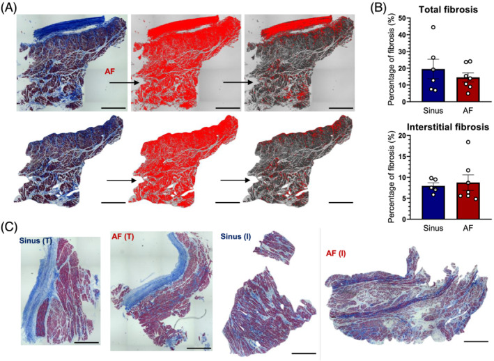
Methods and results: Left atrial samples of 24 male patients with end stage ischemic HF undergoing heart transplantation were analysed. Twelve patients suffered from sustained AF while the others had no documented AF. The expression of inflammasome sensors and their downstream signalling were investigated by Western blot. No differences were observed in the expression of inflammasome sensors between the two groups, while cleaved caspase-1 increased tendentiously in the AF group (P = 0.051). Cleaved caspase-1 also showed significant correlation with the expression of interleukin-1β and its cleaved form in the total population and in the AF group (P < 0.05). The presence of myocardial and epicardial macrophages were assessed by ionized calcium-binding adaptor molecule 1 (Iba1) immunostaining. Number of macrophages showed a tendency towards elevation in the left atrial myocardium and epicardium of AF compared with SR group. The amount of total and interstitial fibrosis was determined on Masson's trichrome-stained sections. Histological assessment revealed no difference between AF and SR groups in the amount of either total or interstitial fibrosis.
Conclusions: This is the first study on inflammation-related differences between HF with SR or AF showing elevated inflammasome activity and enhanced macrophage infiltration in left atrial samples of patients with AF.
Keywords: Atrial fibrillation; Fibrosis; Heart failure; Inflammasome; Macrophages.
© 2022 The Authors. ESC Heart Failure published by John Wiley & Sons Ltd on behalf of European Society of Cardiology.
Conflict of interest statement
P.F. is the founder and CEO of Pharmahungary Group, a group of R&D companies. All other authors have nothing to disclose.
Figures


References
-
- Heijman J, Muna AP, Veleva T, Molina CE, Sutanto H, Tekook M, Wang Q, Abu‐Taha IH, Gorka M, Künzel S, El‐Armouche A, Reichenspurner H, Kamler M, Nikolaev V, Ravens U, Li N, Nattel S, Wehrens XHT, Dobrev D. Atrial myocyte NLRP3/CaMKII nexus forms a substrate for postoperative atrial fibrillation. Circ Res. 2020; 127: 1036–1055. - PMC - PubMed
-
- Onódi Z, Ruppert M, Kucsera D, Sayour AA, Tóth VE, Koncsos G, Novák J, Brenner GB, Makkos A, Baranyai T, Giricz Z, Görbe A, Leszek P, Gyöngyösi M, Horváth IG, Schulz R, Merkely B, Ferdinandy P, Radovits T, Varga ZV. AIM2‐driven inflammasome activation in heart failure. Cardiovasc Res. 2021; 117: 2639–2651. - PubMed
-
- Nessler B, Nessler J, Kitlinski M, Gackowski A, Piwowarska W. Inflammatory markers (TNF‐alpha, IL‐6, CRP), BNP and spiroergometric stress test parameters in patients with heart failure and atrial fibrillation. In 4th Asian Pacific Congress of Heart Failure, Melbourne, Australia. Australia: Elsevier Masson SAS; 2008. Abstract 17S. p S34.
-
- Targoński R, Salczyńska D, Sadowski J, Cichowski L. Relationship between inflammatory markers and clinical patterns of atrial fibrillation in patients with congestive heart failure. Kardiol Pol. 2008; 66: 729–736. - PubMed
Publication types
MeSH terms
Substances
LinkOut - more resources
Full Text Sources
Medical
Research Materials
Miscellaneous
