

The utility of CT virtual bronchoscopy in the esophageal lung diagnosis: A case report
- PMID: 35585904
- PMCID: PMC9108735
- DOI: 10.1016/j.rmcr.2022.101658
The utility of CT virtual bronchoscopy in the esophageal lung diagnosis: A case report
Abstract
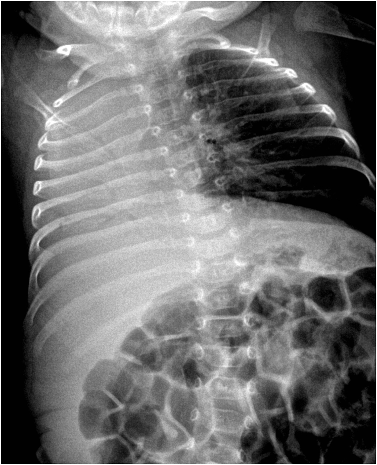
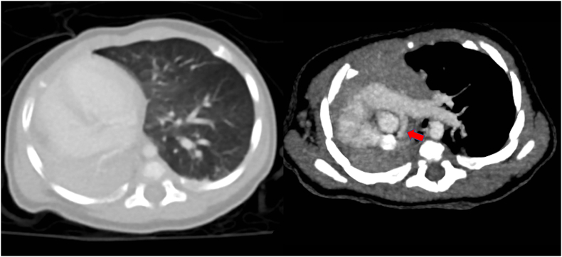
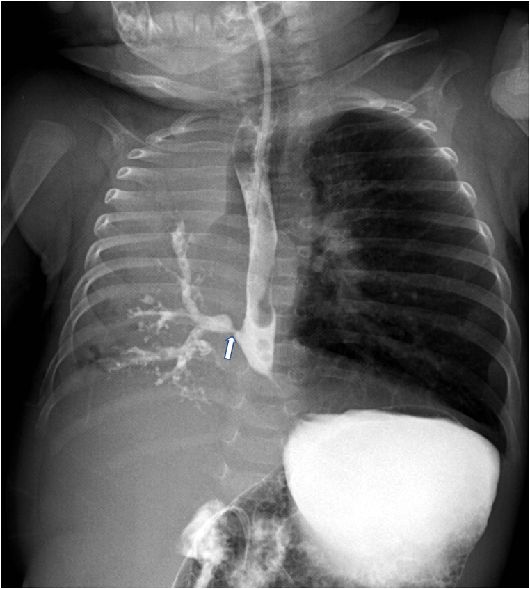
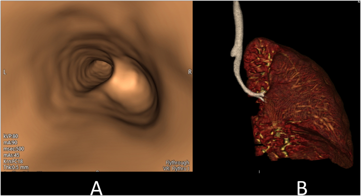
The esophageal lung is a variant of the communicating bronchopulmonary foregut malformation (CBPFM). It needs a high index of suspicion for diagnosis because it is a rare condition and does not have specific symptoms. A CT scan or an esophageal contrast study, showing direct communication between the airways and the esophagus or stomach, confirms the diagnosis. Patients with esophageal lung need flexible bronchoscopy for evaluating tracheobronchial anomalies. We present a three-month-old boy with a right esophageal lung in which the CT virtual bronchoscopy showed an absence of the right main bronchus at the carina level. This case report highlights the importance of CT virtual bronchoscopy as an alternative to flexible bronchoscopy for the diagnosis of tracheobronchial anomalies associated with CBPFM.
Keywords: CBPFM; Communicating bronchopulmonary foregut malformation; Esophageal lung; Tracheoesophageal fistula; Virtual bronchoscopy.
© 2022 The Authors.
Conflict of interest statement
We declare that we have no conflict of interest.
Figures
Similar articles
-
Successful thoracoscopic treatment for tracheoesophageal fistula and esophageal atresia of communicating bronchopulmonary foregut malformation group IB with dextrocardia: a case report of VACTERL association.Surg Case Rep. 2021 Jan 6;7(1):11. doi: 10.1186/s40792-020-01099-y. Surg Case Rep. 2021. PMID: 33409676 Free PMC article.
-
Esophageal lung misdiagnosed as tracheoesophageal fistula: A case report.Radiol Case Rep. 2023 Jan 12;18(3):1227-1231. doi: 10.1016/j.radcr.2022.12.029. eCollection 2023 Mar. Radiol Case Rep. 2023. PMID: 36660583 Free PMC article.
-
Communicating Bronchopulmonary Foregut Malformation Type IB: Diagnostic and Surgical Challenges.European J Pediatr Surg Rep. 2021 Dec 13;9(1):e80-e83. doi: 10.1055/s-0041-1740321. eCollection 2021 Jan. European J Pediatr Surg Rep. 2021. PMID: 34917448 Free PMC article.
-
Congenital bronchopulmonary foregut malformation: systematic review of the literature.BMC Pediatr. 2019 Sep 2;19(1):305. doi: 10.1186/s12887-019-1686-1. BMC Pediatr. 2019. PMID: 31477056 Free PMC article.
-
Communicating bronchopulmonary foregut malformations: classification and embryogenesis.J Pediatr Surg. 1992 Jun;27(6):732-6. doi: 10.1016/s0022-3468(05)80103-4. J Pediatr Surg. 1992. PMID: 1501033 Review.
References
-
- Ballouhey Q., Abbo O., Rouquette I., Rittié J.L., Vial J., Galinier P. Complex communicating bronchopulmonary foregut malformation with pancreatic heterotopy depicted with fetal magnetic resonance imaging: a case report. J. Pediatr. Surg. 2012;47:e7. doi: 10.1016/j.jpedsurg.2011.12.020. - DOI - PubMed
Publication types
LinkOut - more resources
Full Text Sources
