

Structural and Functional Characterization of Gray Matter Alterations in Female Patients With Neuropsychiatric Systemic Lupus
- PMID: 35585919
- PMCID: PMC9108669
- DOI: 10.3389/fnins.2022.839194
Structural and Functional Characterization of Gray Matter Alterations in Female Patients With Neuropsychiatric Systemic Lupus
Abstract
Objective: To investigate morphological and functional alterations within gray matter (GM) in female patients with neuropsychiatric systemic lupus (NPSLE) and to explore their clinical significance.
Methods: 54 female patients with SLE (30 NPSLE and 24 non-NPSLE) and 32 matched healthy controls were recruited. All subjects received a quantitative MRI scan (FLAIR, 3DT1, resting-state functional MRI). GM volume (GMV), fractional amplitude of low-frequency fluctuation (fALFF), regional homogeneity (ReHo), and degree of centrality (DC) were obtained. Between-group comparison, clinical correlation, and discrimination of NPSLE from non-NPSLE were achieved by voxel-based analysis, cerebellar seed-based functional connectivity analysis, regression analysis, and support vector machine (SVM), respectively.
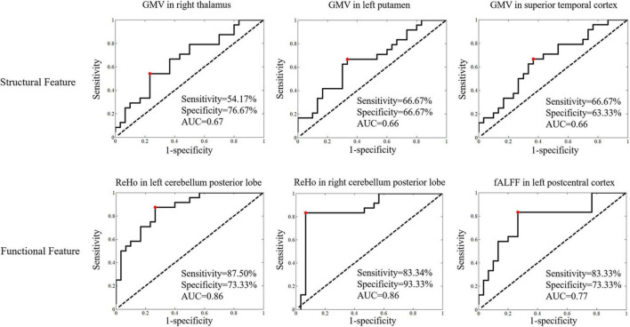
Results: Patients with NPSLE showed overt subcortical GM atrophy without significantly abnormal brain functions in the same region compared with controls. The dysfunction within the left superior temporal gyri (L-STG) was found precede the GM volumetric loss. The function of the nodes in default mode network (DMN) and salience network (SN) were weakened in NPSLE patients compared to controls. The function of the cerebellar posterior lobes was significantly activated in non-NPSLE patients but attenuated along with GM atrophy and presented higher connectivity with L-STG and DMN in NPSLE patients, while the variation of the functional activities in the sensorimotor network (SMN) was the opposite. These structural and functional alterations were mainly correlated with disease burden and anti-phospholipid antibodies (aPLs) (r ranges from -1.53 to 1.29). The ReHos in the bilateral cerebellar posterior lobes showed high discriminative power in identifying patients with NPSLE with accuracy of 87%.
Conclusion: Patients with NPSLE exhibit both structural and functional alterations in the GM of the brain, which especially involved the deep GM, the cognitive, and sensorimotor regions, reflecting a reorganization to compensate for the disease damage to the brain which was attenuated along with pathologic burden and cerebral vascular risk factors. The GM within the left temporal lobe may be one of the direct targets of lupus-related inflammatory attack. The function of the cerebellar posterior lobes might play an essential role in compensating for cortical functional disturbances and may contribute to identifying patients with suspected NPSLE in clinical practice.
Keywords: cerebellar seed-based functional connectivity; female; gray matter; neurosychiatric systemic lupus; resting sate fMRI.
Copyright © 2022 Su, Zhuo, Duan, Huang, Qiu, Li, Liu and Zeng.
Conflict of interest statement
The authors declare that the research was conducted in the absence of any commercial or financial relationships that could be construed as a potential conflict of interest.
Figures


Similar articles
-
Alterations of spontaneous brain activity in systematic lupus erythematosus patients without neuropsychiatric symptoms: A resting-functional MRI study.Lupus. 2021 Oct;30(11):1781-1789. doi: 10.1177/09612033211033984. Epub 2021 Oct 8. Lupus. 2021. PMID: 34620007
-
Abnormal amplitude of low frequency fluctuation and functional connectivity in non-neuropsychiatric systemic lupus erythematosus: a resting-state fMRI study.Neuroradiology. 2019 Mar;61(3):331-340. doi: 10.1007/s00234-018-2138-6. Epub 2019 Jan 12. Neuroradiology. 2019. PMID: 30637462
-
Altered Functional Brain Network in Systemic Lupus Erythematosus Patients Without Overt Neuropsychiatric Symptoms Based on Resting-State Functional Magnetic Resonance Imaging and Multivariate Pattern Analysis.Front Neurol. 2021 Jul 21;12:690979. doi: 10.3389/fneur.2021.690979. eCollection 2021. Front Neurol. 2021. PMID: 34354663 Free PMC article.
-
Different patterns of cerebral perfusion in SLE patients with and without neuropsychiatric manifestations.Hum Brain Mapp. 2020 Feb 15;41(3):755-766. doi: 10.1002/hbm.24837. Epub 2019 Oct 24. Hum Brain Mapp. 2020. PMID: 31650651 Free PMC article.
-
Advanced neuroimaging in neuropsychiatric systemic lupus erythematosus.Curr Opin Neurol. 2020 Jun;33(3):353-361. doi: 10.1097/WCO.0000000000000822. Curr Opin Neurol. 2020. PMID: 32349105 Free PMC article. Review.
Cited by
-
Cerebral network topology and peak width of skeletonized mean diffusivity changes associated with cognitive impairment in patients with obstructive sleep apnea.Brain Imaging Behav. 2025 Aug 15. doi: 10.1007/s11682-025-01045-2. Online ahead of print. Brain Imaging Behav. 2025. PMID: 40813499
-
Serotonin Syndrome-Mimicking Manifestations in a Patient with Systemic Lupus Erythematosus.J Clin Med. 2024 Jun 15;13(12):3516. doi: 10.3390/jcm13123516. J Clin Med. 2024. PMID: 38930045 Free PMC article.
-
Abnormal brain functional networks in systemic lupus erythematosus: a graph theory, network-based statistic and machine learning study.Brain Commun. 2025 Apr 2;7(2):fcaf130. doi: 10.1093/braincomms/fcaf130. eCollection 2025. Brain Commun. 2025. PMID: 40207059 Free PMC article.
-
MRI-based neuroimaging alterations in immune-related skin diseases: a comprehensive review.Arch Dermatol Res. 2025 Mar 8;317(1):529. doi: 10.1007/s00403-025-04023-2. Arch Dermatol Res. 2025. PMID: 40056246 Review.
-
Cerebral Microstructural and Microvascular Changes in Non-Neuropsychiatric Systemic Lupus Erythematosus: A Study Using Diffusion Kurtosis Imaging and 3D Pseudo-Continuous Arterial Spin Labeling.J Inflamm Res. 2023 Nov 22;16:5465-5475. doi: 10.2147/JIR.S429521. eCollection 2023. J Inflamm Res. 2023. PMID: 38026250 Free PMC article.
References
-
- Barraclough M., McKie S., Parker B., Jackson A., Pemberton P., Elliott R., et al. (2019). Altered cognitive function in systemic lupus erythematosus and associations with inflammation and functional and structural brain changes. Ann. Rheum. Dis. 78 934–940. 10.1136/annrheumdis-2018-214677 - DOI - PMC - PubMed
LinkOut - more resources
Full Text Sources