. 2022 May 2:16:860280.
doi: 10.3389/fnins.2022.860280. eCollection 2022.

Dietary Supplementation With Acer truncatum Oil Promotes Remyelination in a Mouse Model of Multiple Sclerosis

Affiliations

Dietary Supplementation With Acer truncatum Oil Promotes Remyelination in a Mouse Model of Multiple Sclerosis

Yuhuan Xue et al. Front Neurosci. .

Abstract

Background: Multiple sclerosis is a chronic demyelinating disease of uncertain etiology. Traditional treatment methods produce more adverse effects. Epidemiological and clinical treatment findings showed that unknown environmental factors contribute to the etiology of MS and that diet is a commonly assumed factor. Despite the huge interest in diet expressed by people with MS and the potential role diet plays in MS, very little data is available on the role of diet in MS pathogenesis and MS course, in particular, studies on fats and MS. The oil of Acer truncatum is potential as a resource to be exploited in the treatment of some neurodegenerative diseases.

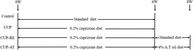
Objective: Here, we investigated the underlying influences of Acer truncatum oil on the stimulation of remyelination in a cuprizone mouse model of demyelination.

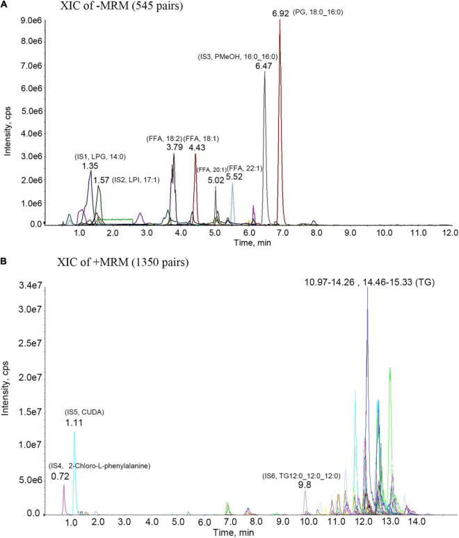
Methods: Cuprizone (0.2% in chow) was used to establish a mouse model of demyelination. Acer truncatum oil was administrated to mice during remyelination. Following techniques were used: behavioral test, histochemistry, fluorescent immunohistochemistry, transmission electron microscope.

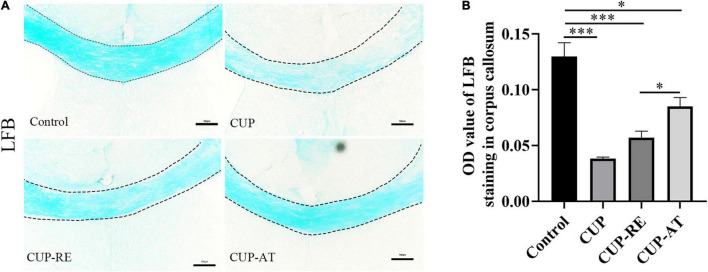
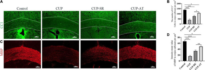
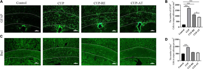
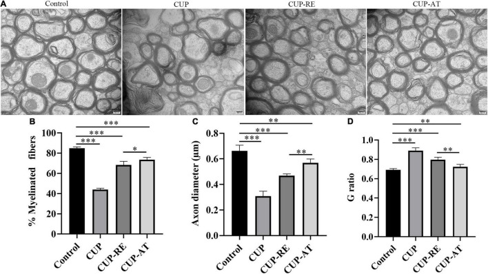
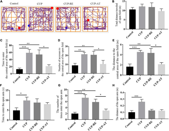
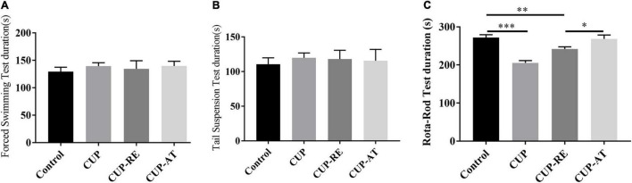
Results: Mice exposed to cuprizone for 6 weeks showed schizophrenia-like behavioral changes, the increased exploration of the center in the open field test (OFT), increased entries into the open arms of the elevated plus-maze, as well as demyelination in the corpus callosum. After cuprizone withdrawal, the diet therapy was initiated with supplementation of Acer truncatum oil for 2 weeks. As expected, myelin repair was greatly enhanced in the demyelinated regions with increased mature oligodendrocytes (CC1) and myelin basic protein (MBP). More importantly, the supplementation with Acer truncatum oil in the diet reduced the schizophrenia-like behavior in the open field test (OFT) and the elevated plus-maze compared to the cuprizone recovery group. The results revealed that the diet supplementation with Acer truncatum oil improved behavioral abnormalities, oligodendrocyte maturation, and remyelination in the cuprizone model during recovery.

Conclusion: Diet supplementation with Acer truncatum oil attenuates demyelination induced by cuprizone, indicating that Acer truncatum oil is a novel therapeutic diet in demyelinating diseases.

Keywords: Acer truncatum oil; cuprizone; demyelination; neurodegenerative disease; remyelination.


Conflict of interest statement

The authors declare that the research was conducted in the absence of any commercial or financial relationships that could be construed as a potential conflict of interest.

Figures

FIGURE 1
FIGURE 1
Experimental time course image.
FIGURE 2
FIGURE 2
MRM metabolite assay multi-peak chart. IS1-IS6 are all internal standard; (A)-MRM metabolite assay multi-peak chart. 3.79- (FFA, 18:2)- Linoleic acid; 4.43- (FFA, 18:1)- Oleic acid; 5.02- (FFA, 20:1)- Eicosenoic Acid; 5.52- (FFA, 22:1)- Erucic acid; 6.92- (PG, 18:0_16:0)- Phosphatidyl Glycerols (18:0_16:0); (B) + MRM metabolite assay multi-peak chart. 11.33- (TG,16:0_18:2_18:2)- Triglyceride (16:0_18:2_18:2); 11.7- (TG,18:2_18:2_20:1)- Triglyceride (18:2_18:2_20:1); 12.15- (TG, 18:2_18:2_22:1)- Triglyceride (18:2_18:2_22:1); 13- (TG,18:1_18:1_22:1)- Triglyceride (18:1_18:1_22:1).
FIGURE 3
FIGURE 3
Administration of CUP induced demyelination of the corpus callosum and dietary Acer truncatum oil promoted remyelination in the corpus callosum. (A) LFB staining in the corpus callosum. The corpus callosum in control group was densely and regularly labeled with LFB, whereas the LFB staining in mice administrated with CUP became light and irregular. After dietary Acer truncatum oil, the LFB staining recovered almost like that in control group. (B) Quantitative analysis of OD value of LFB in the corpus callosum. Acute CUP administration significantly reduces the OD value of LFB. Dietary Acer truncatum oil rescued the reduction of OD value of LFB. n = 3–4/group. Scale bar: 100 μm. Data were expressed as means ± SEM. Differences between groups were expressed as *p < 0.05, ***p < 0.001.
FIGURE 4
FIGURE 4
CUP affects the maturation of oligodendrocytes in the corpus callosum, supplementation with Acer truncatum oil accelerated the maturation of oligodendrocytes and promoted remyelination. (A) CC1 immunofluorescence staining was performed in coronal slices of the corpus callosum in different groups. In control group, the CC1-positive mature oligodendrocytes in the corpus callosum were densely arranged, whereas the number of mature oligodendrocytes in the corpus callosum administrated with CUP significantly reduced. (B) Quantitative analysis of CC1-positive cells in the corpus callosum. The density of CC1-positive cells was significantly reduced in the CUP group. Dietary Acer truncatum oil significantly increased the number of CC1-positive cells. n = 4–6/group. (C) Fine MBP-positive fibers were well observed in the corpus callosum from control mice. In contrast, the mice exposed to CUP for 6 weeks showed widespread myelin breakdown. After dietary Acer truncatum oil, the MBP immunofluorescence staining recovered almost like that in control group. (D) Quantitative analysis of fluorescence intensity means the value of MBP in the corpus callosum. MBP expression in the corpus callosum was significantly decreased after CUP exposure, whereas Acer truncatum oil supplementation significantly increased MBP expression. n = 8/group. Scale bar: 100 μm. Data were expressed as means ± SEM. Differences between groups were expressed as *p < 0.05, **p < 0.01, ***p < 0.001.
FIGURE 5
FIGURE 5
Supplementation with Acer truncatum oil inhibited the activation of microglia and astrocytes after administration with CUP. (A) GFAP immunofluorescent staining (green) was performed in coronal slices of the corpus callosum. In control mice, astrocytes were only sporadically observed. CUP induced an increase in the number of astrocytes and cytosolic hypertrophy, which were attenuated by Acer truncatum oil supplementation. (B) Quantitative analysis of GFAP-positive cells in the corpus callosum. CUP administration significantly increases the number of astrocytes, dietary supplementation of Acer truncatum oil significantly decreased the number of GFAP-positive cells. n = 4/group. (C) Iba1 immunofluorescent staining (green) was performed in coronal slices of the corpus callosum. In control mice, microglia were only sporadically observed. CUP induced an increase in the number of microglia, which were attenuated by Acer truncatum oil supplementation. (D) Quantitative analysis of Iba1-positive cells in the corpus callosum. CUP administration significantly increased the number of astrocytes, supplementation of Acer truncatum oil decreased the number of astrocytes cells. However, the difference was not significant. n = 4–6/group. Data were expressed as means ± SEM. Differences between groups were expressed as *p < 0.05, ***p < 0.001.
FIGURE 6
FIGURE 6
Acer truncatum oil supplementation can be of advantage to recover the damage of axonal and myelin in the MS mouse model. (A) The TEM photomicrographs were taken from coronal sections of the corpus callosum, healthy myelin sheaths with normal and intact structures were observed of the corpus callosum in control group, whereas disintegration of neural fibers was found in CUP mice. Acer truncatum oil supplementation was efficient in the recovery of myelin sheaths from pathological morphology, in comparison to CUP-RE group mice, in which lower electron density and less well-defined myelin sheaths was found. (B) Quantitative analysis of the myelinated fibers. The percentage of the myelinated fibers in the corpus callosum of mice administrated with CUP significantly decreased compared with that in control group. Dietary Acer truncatum oil supplementation significantly increased in axon diameter as compared to that in the CUP-RE group. (C) Quantitative analysis of the axon diameter. Axon diameter in mice administrated with CUP significantly decreased compared with that in control group, Axon diameter after dietary Acer truncatum oil supplementation significantly increased as compared to that in the CUP-RE group. (D) Quantitative analysis of the G ratio. The G ratio in CUP mice was significantly increased compared with that in control group, after dietary Acer truncatum oil treatment, the G ratio was significantly reduced compared with the CUP-RE group. n = 9/group. Scale bar: 200 nm. Data were expressed as means ± SEM. Differences between groups were expressed as *p < 0.05, **p < 0.01, ***p < 0.001.
FIGURE 7
FIGURE 7
Dietary supplementation of Acer truncatum oil promoted the recovery of CUP-induced behavioral changes in mice. (A) Representative trajectory images of each group of OFT. (B–E) No significant difference in the total distance was found in the OFT between the groups. The activity time (C), number (D) and accumulated distance (E) of the CUP-exposed mice in the central area of the OFT were significantly higher than that of mice in control group; After CUP-exposure, 2-week Acer truncatum oil dietary supplementation promoted the recovery of CUP-induced behavioral changes compared to that in mice of CUP-RE group. n = 8–12/group. (F–H) CUP-exposed mice showed significantly higher activity time (F) number (G) and distance traveled (H) in the open arm of EMP. Dietary supplementation of Acer truncatum oil significantly reduced the number of mice entering the open arm compared with that in CUP-RE. n = 6–7/group. Data were expressed as means ± SEM. Differences between groups were expressed as *p < 0.05, **p < 0.01, ***p < 0.001.
FIGURE 8
FIGURE 8
Dietary supplementation of Acer truncatum oil improved the recovery of CUP-induced physical coordination in mice. (A) The immobility time of the last 4 min of the 5 min of FST. n = 8–15/group. (B) The immobility time of the last 4 min of the 5 min of TST. n = 6–10/group. (C) Dietary supplementation of Acer truncatum oil significantly improved the recovery of CUP-induced Rota-rod changes in mice. n = 8–12/group. Data were expressed as means ± SEM. Differences between groups were expressed as *p < 0.05, **p < 0.01, ***p < 0.001.

Similar articles

Cited by

References

    1. Abulimiti A., Teng G., Jue H., Kelly H., Wenqiang W., Xinmin L. (2015). The protective effects of areca catechu extract on cognition and social interaction deficits in a Cuprizone-induced demyelination model. Evid. Based Complement. Alternat. Med. 2015:426092. - PMC - PubMed
    1. Alves-Bezerra M., Cohen D. E. (2017). Triglyceride metabolism in the liver. Compr. Physiol. 8 1–8. 10.1002/cphy.c170012 - DOI - PMC - PubMed
    1. Alter M., Yamoor M., Harshe M. (1974). Multiple sclerosis and nutrition. Arch. Neurol. 31, 267–72. - PubMed
    1. Armand M., Borel P., Pasquier B., Dubois C., Senft M., Andre M., et al. (1996). Physiological characteristics of emulsions during fat digestion in human stomach and duodenum. Am. J. Physiol. 271 G172–G183. 10.1152/ajpgi.1996.271.1.G172 - DOI - PubMed
    1. Ascherio A., Munger K. L., Lünemann J. D. (2012). The initiation and prevention of multiple sclerosis. Nat. Rev. Neurol. 8 602–612. 10.1038/nrneurol.2012.198 - DOI - PMC - PubMed