. 2022 May 2:13:872047.
doi: 10.3389/fimmu.2022.872047. eCollection 2022.

Furin and TMPRSS2 Resistant Spike Induces Robust Humoral and Cellular Immunity Against SARS-CoV-2 Lethal Infection

Affiliations

Furin and TMPRSS2 Resistant Spike Induces Robust Humoral and Cellular Immunity Against SARS-CoV-2 Lethal Infection

Jhe-Jhih Lin et al. Front Immunol. .

Abstract

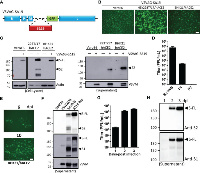
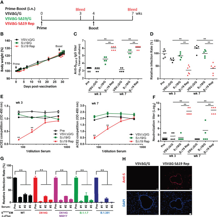
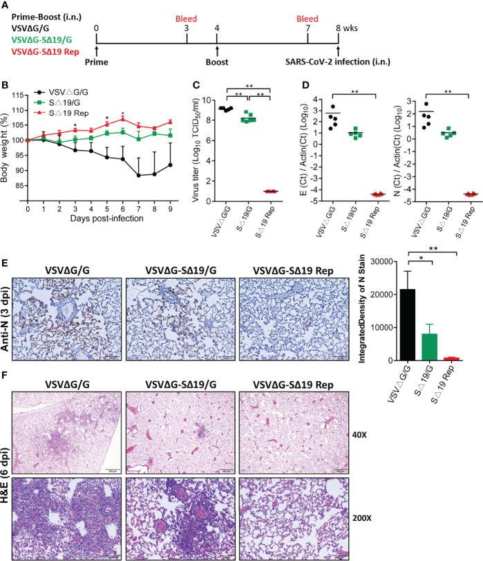
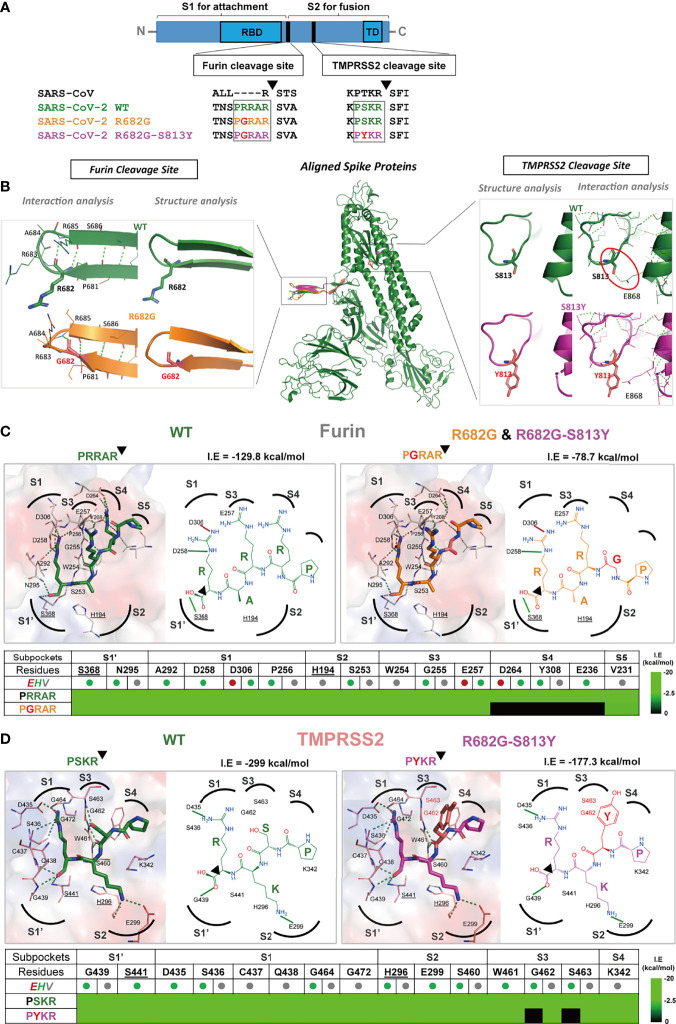
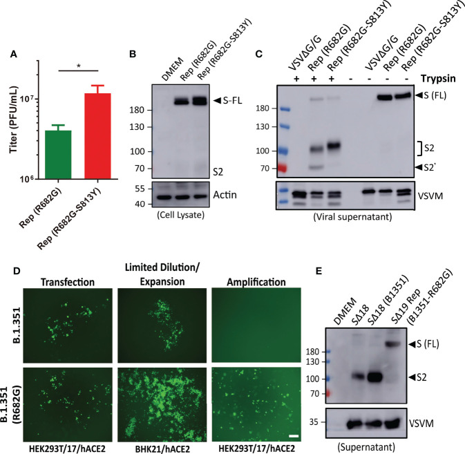
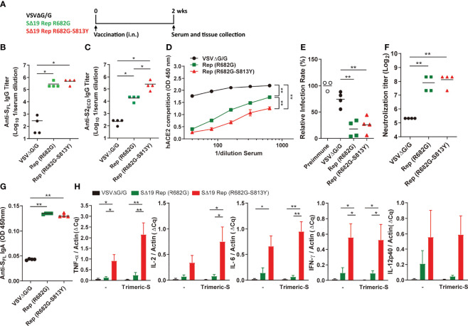
An effective COVID-19 vaccine against broad SARS-CoV-2 variants is still an unmet need. In the study, the vesicular stomatitis virus (VSV)-based vector was used to express the SARS-CoV-2 Spike protein to identify better vaccine designs. The replication-competent of the recombinant VSV-spike virus with C-terminal 19 amino acid truncation (SΔ19 Rep) was generated. A single dose of SΔ19 Rep intranasal vaccination is sufficient to induce protective immunity against SARS-CoV-2 infection in hamsters. All the clones isolated from the SΔ19 Rep virus contained R682G mutation located at the Furin cleavage site. An additional S813Y mutation close to the TMPRSS2 cleavage site was identified in some clones. The enzymatic processing of S protein was blocked by these mutations. The vaccination of the R682G-S813Y virus produced a high antibody response against S protein and a robust S protein-specific CD8+ T cell response. The vaccinated animals were protected from the lethal SARS-CoV-2 (delta variant) challenge. The S antigen with resistance to enzymatic processes by Furin and TMPRSS2 will provide better immunogenicity for vaccine design.

Keywords: ACE2 transgenic mice; S1/S2 cleavage site; SARS-CoV-2 Spike; TMPRSS2; VSV; furin; pseudotype; replication-competent.


Conflict of interest statement

The authors declare that the research was conducted in the absence of any commercial or financial relationships that could be construed as a potential conflict of interest.

Figures

Figure 1
Figure 1
Generation of recombinant vesicular stomatitis virus (VSV) expressing the C-terminal 19 amino acid deletion SARS-CoV-2 spike mutant (VSVΔG-SΔ19) with replication capability. (A) Schematic representation of the genomic organization of the G protein-deficient VSV vector (VSVΔG) inserted with the C-terminal 19 amino acid truncated SARS-CoV-2 spike protein (SΔ19). N, nucleoprotein; P, phosphoprotein; M, matrix; GFP, green fluorescent protein; L, large polymerase. (B, C) Vero E6, HEK293T-hACE2, and BHK21-hACE2 cells were infected with the VSVΔG-SΔ19 virus (early passage, moi=0.5). Virus-driven GFP expression was monitored by fluorescence microscopy at 24 hrs post-infection (B). The SΔ19 protein expression in cells (left panel) and supernatant (right panel) was examined by immunoblotting (C). (D) The VSVΔG-SΔ19 virus was propagated in HEK293T-hACE2 cells, and the virus titer was determined in BHK21-hACE2 cells. (E) After a few passages in HEK293T-hACE2 cells, the viruses were inoculated in BHK21-hACE2 cells with limiting dilution. The replication-competent VSVΔG-SΔ19 viruses (SΔ19 Rep) emerging from BHK21-hACE2 cells were observed with GFP monitoring. (F) The S protein expression on recombinant virus particles of VSVΔG/G, the early passage of VSVΔG-SΔ19 enveloped with the VSV glycoprotein (SΔ19/G; replication incompetent) and SΔ19 Rep virus was examined by immunoblotting. (G, H) Vero E6 cells were infected with the SΔ19 Rep virus (moi=1, n=3). Virus titer (G) and S protein expression on virus particles (H) in culture supernatant were examined. moi, multiplicity of infection; dpi, days post-infection. Scale bar: 100 µm.
Figure 2
Figure 2
The SΔ19 Rep virus effectively stimulates anti-spike antibodies with neutralizing activity in hamsters. (A) Experimental design of the prime-boost vaccination with VSVΔG/G (control virus), VSVΔG-SΔ19/G (replication-incompetent virus), and SΔ19 Rep in Gold Syrian hamster by intranasal (i.n.) administration (1×10⁶ pfu or ffu/hamster; n=5-6). Body weight of vaccinated hamsters was monitored periodically (B). Serum samples collected from 3 and 7 weeks post-vaccination were subjected to ELISA for anti-spike specific IgG antibody (C), the VSVΔG-Spike pseudovirus-based neutralization assay (D), the RBD-hACE2 interaction competition assay (E), the SARS-CoV-2 neutralization assay (F), and the the Svariant EM-LvFluc viruses-based neutralization assay (G). (H) Lung sections obtained from hamsters infected with VSVΔG/G or SΔ19 Rep (1×10⁶ pfu or ffu/hamster; i.n.) at 20 hrs post-infection were stained with anti-S antibodies and counterstained with DAPI. **P < 0.01. Scale bar: 20 μm.
Figure 3
Figure 3
Prime-boost SΔ19 Rep vaccination protects hamsters from SARS-CoV-2 infection. (A) Experimental design of the prime-boost vaccination and SARS-CoV-2 challenge (1×10⁵ TCID50/hamster; i.n.) in hamsters. (B) Body weight loss post-SARS-CoV-2 challenge. (C) The virus titers of SARS-CoV-2 in the lung at 3 dpi were detected by TCID50 assay. (D) Viral RNA transcripts of E and N genes in the lung (3 dpi) were measured by quantitative real-time PCR. (E) Viral N protein expression in the lung (3 dpi) was detected by immunohistochemistry and quantified using ImageJ software. Scale bar: 100 μm. (F) H&E staining of lung tissue at 6 dpi. Scale bar: 500 μm (40X) and 100 μm (200X). *P < 0.05; **P < 0.01.
Figure 4
Figure 4
A single dose SΔ19 Rep vaccination induces effective immunity against SARS-CoV-2 infection. (A) Experimental design of a single dose vaccination (1×10⁶ pfu or ffu/hamster; i.n.; n=10) and SARS-CoV-2 challenge (1×10⁵ TCID50/hamster; i.n.). (B) Body weight loss post-SARS-CoV-2 challenge. SARS-CoV-2 virus titer (C), viral RNA (D), and N protein expression (E) in the lung at 3 dpi were assessed. Scale bar: 20 μm. (F) H&E staining of lung tissue (3, 6, and 9 dpi). Scale bar: 500 μm (40X) and 100 μm (200X). *P < 0.05; **P < 0.01.
Figure 5
Figure 5
The R682G and S813Y mutations in Spike protein lead to altered enzyme binding and proteolysis. (A) The Furin and TMPRSS2 cleavage motifs are shown in the spike protein sequences of SARS-CoV, SARS-CoV-2 WT, and mutants. Furin cleavage occurs between the S1 and S2 subunits, while the TMPRSS2 cleavage occurs within the S2 subunit. (B) The S protein structure and interactions of the WT (wild-type; green), R682G (orange), and R682G-S813Y (magenta) mutants were modeled using the Phyre2 web portal. The full spike proteins are aligned, and the specific mutation sites are shown in insights of the Furin and TMPRSS2 cleavage sites. (C, D) Molecular docking of spike protein peptide substrates (of WT and mutants) with Furin and TMPRSS2 using iGEMDOCK. The protease active site (surface) and the binding poses (2D diagrams) of the WT (green stick) and mutants (mutated residue in red sticks) were shown. The active site subpockets (dotted curves) with residues (catalytic residues labels underlined) were also displayed. The interactions (solid black lines, E-dark red, H-green, V-grey) and total interaction energies (I.E in kcal/mol) were examined. The interaction table details the substrate-subpocket residue interactions.
Figure 6
Figure 6
R682G and S813Y mutations facilitate rVSV-S replication. (A) HEK293T-hACE2 cells were infected with the SΔ19 Rep virus with R682G or R682G-S813Y mutation. Virus titer in the culture supernatant was measured (moi=0.1, 3 dpi, n=4). (B) Virus-infected cell lysate (moi=1, 1 dpi) was subjected to immunoblotting with anti-S2 antibodies. (C) The viral particles in the culture supernatant were digested with TPCK-trypsin (2 μg/ml at 37°C for 30 min) and analyzed by immunoblotting. (D) SΔ19 beta variant (B.1.351) gene with and without R682G mutation was inserted into the VSVΔG-GFP DNA vector and co-transfected with all the required helper plasmids in HEK293T-hACE2 cells to rescue recombinant VSVΔG-SΔ19 (B.1.351)/G virus. Virus replication was monitored by GFP expression. The rescued viruses were collected to infect BHK21-hACE2 cells with limiting dilution, and further amplified in HEK293T-hACE2 cells. (E) The Spike protein expression pattern of the replication-competent SΔ19 (B.1.351-R682G) virus was examined by immunoblotting alone with SΔ18 and SΔ18 (B.1.351) pseudoviruses for comparison. Scale bar: 100 μm. *P < 0.05.
Figure 7
Figure 7
The SΔ19 Rep (R682G-S813Y) mutant stimulates robust neutralizing antibodies and Th1 immune response in hamsters. (A) Experimental design of a single dose vaccination with VSVΔG/G (control virus), SΔ19 Rep-R682G and SΔ19 Rep-R682G-S813Y in hamster (i.n.; 1×10⁶ pfu or ffu/hamster; n=4). The serum, spleen, and lung of the vaccinated hamsters were collected 2 weeks post-vaccination. Serum samples were subjected to anti-SFL (B) and anti-S2ECD (C) IgG ELISA, the RBD-hACE2 interaction competition assay (D), and the neutralization assay with the VSVΔG-based S pseudotyped virus (E) and SARS-CoV-2 virus (F). The anti-SFL IgA expression in the lung homogenates was measured by ELISA (G). The isolated hamster splenocytes were stimulated with or without trimeric-S protein (5 μg/ml) for 72 hrs, and subjected to detect the TNF-α, IL-2, IL-6, IFN-γ and IL-12 mRNA expression by qRT-PCR (H). *P < 0.05; **P < 0.01.
Figure 8
Figure 8
The SΔ19 Rep (R682G-S813Y)-vaccinated hACE2 transgenic mice were protected from the SARS-CoV-2 lethal infection. (A) K18-hACE2 transgenic mice were vaccinated with VSVΔG/G (control virus), SΔ19 Rep-R682G, and SΔ19 Rep-R682G-S813Y twice on Day 0 and Day 21 (i.n.; 1×10⁸ pfu or ffu/mouse; n=5). Anti-SFL IgG titer in the serum samples collected on Day 14 and Day 28 was measured by ELISA. (B) Anti-SFL IgA titer in Nasal lavage fluid (Day 35) was detected by ELISA. (C–F) Splenocytes were isolated on Day 35-post vaccination and stained with T cell markers and the H-2K(b) SARS-CoV-2 S Tetramer for flow cytometry. Some splenocytes were treated with PMA (50 ng/ml) and ionomycin (500 ng/ml) for 5 h to activate T cell (P+I). (G–J) The vaccinated-mice were challenged with the SARS-CoV-2 Delta strain (1000 TCID50/mouse; i.n.; n=6-8). Mouse body weight loss (G) and survival rate (H) were monitored. Half of the infected-mice were sacrificed, and the virus titer (I) and viral RNA level (J) in the mouse lung were measured. *P < 0.05; **P < 0.01.

Similar articles

Cited by

References

    1. Zheng J. SARS-CoV-2: An Emerging Coronavirus That Causes a Global Threat. Int J Biol Sci (2020) 16:1678–85. doi: 10.7150/ijbs.45053 - DOI - PMC - PubMed
    1. Hoffmann M, Kleine-Weber H, Schroeder S, Kruger N, Herrler T, Erichsen S, et al. . SARS-CoV-2 Cell Entry Depends on ACE2 and TMPRSS2 and Is Blocked by a Clinically Proven Protease Inhibitor. Cell (2020) 181:271–80. doi: 10.1016/j.cell.2020.02.052 - DOI - PMC - PubMed
    1. Xiong X, Qu K, Ciazynska KA, Hosmillo M, Carter AP, Ebrahimi S, et al. . A Thermostable, Closed SARS-CoV-2 Spike Protein Trimer. Nat Struct Mol Biol (2020) 27:934–41. doi: 10.1038/s41594-020-0478-5 - DOI - PMC - PubMed
    1. Walls AC, Park YJ, Tortorici MA, Wall A, McGuire AT, Veesler D. Structure, Function, and Antigenicity of the SARS-CoV-2 Spike Glycoprotein. Cell (2020) 181:281+. doi: 10.1016/j.cell.2020.02.058 - DOI - PMC - PubMed
    1. Huang Y, Yang C, Xu XF, Xu W, Liu SW. Structural and Functional Properties of SARS-CoV-2 Spike Protein: Potential Antivirus Drug Development for COVID-19. Acta Pharmacol Sin (2020) 41:1141–9. doi: 10.1038/s41401-020-0485-4 - DOI - PMC - PubMed

Publication types

Supplementary concepts