. 2022 Apr 27:13:856033.
doi: 10.3389/fimmu.2022.856033. eCollection 2022.

Seroreactivity of the Severe Acute Respiratory Syndrome Coronavirus 2 Recombinant S Protein, Receptor-Binding Domain, and Its Receptor-Binding Motif in COVID-19 Patients and Their Cross-Reactivity With Pre-COVID-19 Samples From Malaria-Endemic Areas

Affiliations

Seroreactivity of the Severe Acute Respiratory Syndrome Coronavirus 2 Recombinant S Protein, Receptor-Binding Domain, and Its Receptor-Binding Motif in COVID-19 Patients and Their Cross-Reactivity With Pre-COVID-19 Samples From Malaria-Endemic Areas

Abdouramane Traoré et al. Front Immunol. .

Abstract

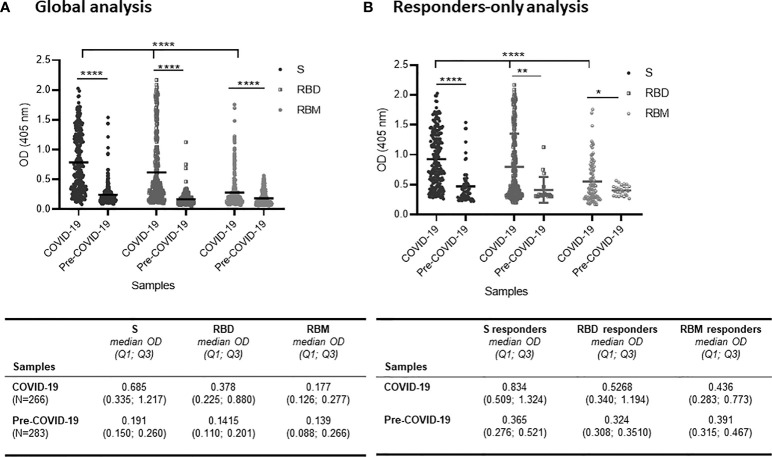
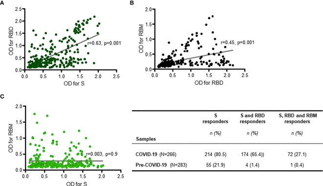
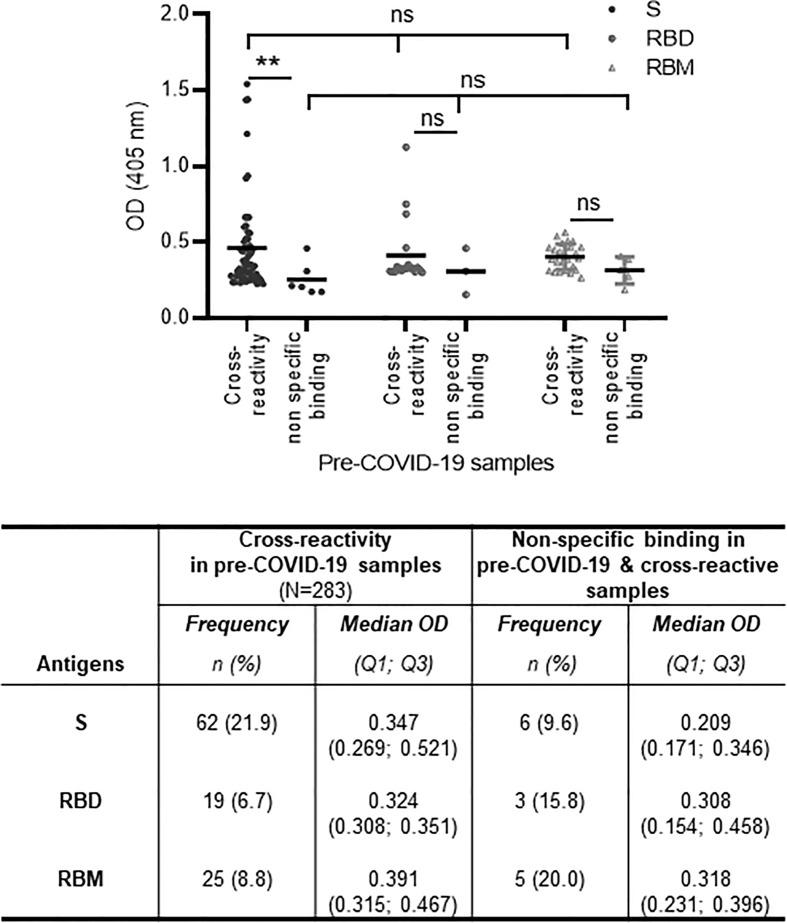
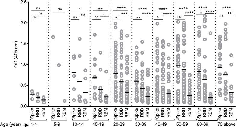
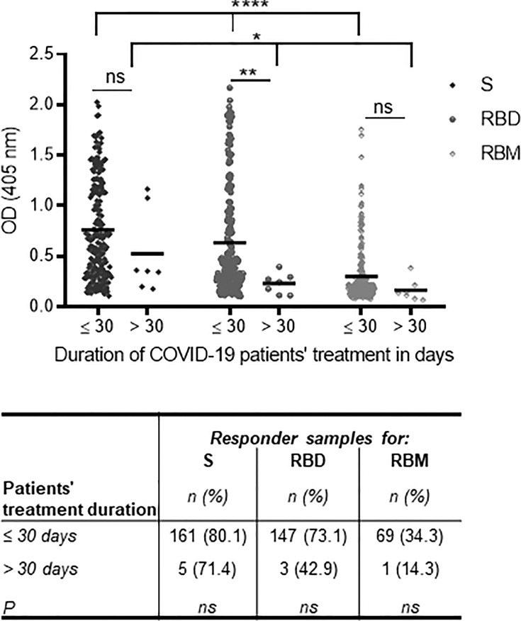
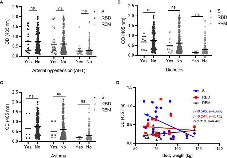
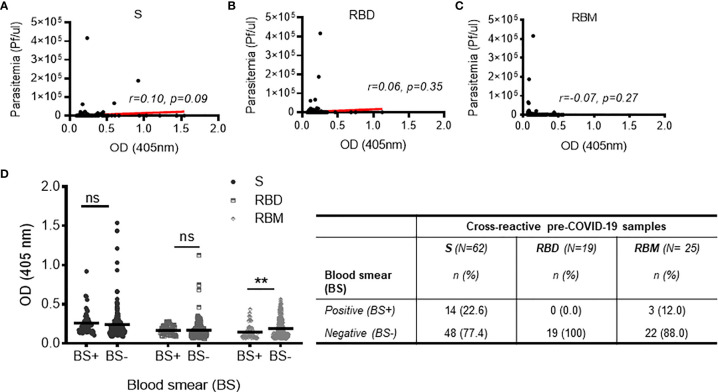
Despite the global interest and the unprecedented number of scientific studies triggered by the COVID-19 pandemic, few data are available from developing and low-income countries. In these regions, communities live under the threat of various transmissible diseases aside from COVID-19, including malaria. This study aims to determine the severe acute respiratory syndrome coronavirus 2 (SARS-CoV-2) seroreactivity of antibodies from COVID-19 and pre-COVID-19 samples of individuals in Mali (West Africa). Blood samples from COVID-19 patients (n = 266) at Bamako Dermatology Hospital (HDB) and pre-COVID-19 donors (n = 283) from a previous malaria survey conducted in Dangassa village were tested by ELISA to assess IgG antibodies specific to the full-length spike (S) protein, the receptor-binding domain (RBD), and the receptor-binding motif (RBM436-507). Study participants were categorized by age, gender, treatment duration for COVID-19, and comorbidities. In addition, the cross-seroreactivity of samples from pre-COVID-19, malaria-positive patients against the three antigens was assessed. Recognition of the SARS-CoV-2 proteins by sera from COVID-19 patients was 80.5% for S, 71.1% for RBD, and 31.9% for RBM (p < 0.001). While antibody responses to S and RBD tended to be age-dependent, responses to RBM were not. Responses were not gender-dependent for any of the antigens. Higher antibody levels to S, RBD, and RBM at hospital entry were associated with shorter treatment durations, particularly for RBD (p < 0.01). In contrast, higher body weights negatively influenced the anti-S antibody response, and asthma and diabetes weakened the anti-RBM antibody responses. Although lower, a significant cross-reactive antibody response to S (21.9%), RBD (6.7%), and RBM (8.8%) was detected in the pre-COVID-19 and malaria samples. Cross-reactive antibody responses to RBM were mostly associated (p < 0.01) with the absence of current Plasmodium falciparum infection, warranting further study.

Keywords: COVID-19 samples; Pre-COVID-19 samples; SARS-CoV-2 S protein; cross-reactivity; malaria endemic-area; seroreactivity.


Conflict of interest statement

The authors declare that the research was conducted in the absence of any commercial or financial relationships that could be construed as a potential conflict of interest.

Figures

Figure 1
Figure 1
Distribution of antibody responses against S, receptor-binding domain (RBD), and receptor-binding motif (RBM) antigens in COVID-19 and pre-COVID-19 samples. (A) Global analysis of samples (positive and negative in ELISA) shows that antibody (Ab) levels (mean OD shown as a horizontal black line in the dot plots) for S, RBD, and RBM were significantly higher in COVID-19 patient samples as compared to pre-COVID-19 donor samples (p < 0.0001). Also, the Ab levels varied significantly (p < 0.0001) among S, RBD, and RBM in COVID-19 samples. The table shows the median OD, Q1, and Q3 values of antibodies for S, RBD, and RBM in COVID-19 and pre-COVID-19 samples. (B) Levels of Ab responses in responder-only COVID-19 samples were significantly higher than in responder-only pre-COVID-19 samples (cross-reactive responders) for S (p < 0.0001), RBD (p < 0.01), and RBM (p < 0.05). The table shows the median OD, Q1, and Q3 of antibodies for S, RBD, and RBM of responder-only samples in COVID-19 and pre-COVID-19 participants. The unpaired t-test and ANOVA were performed to compare the mean ODs of antibodies between the two groups and within the groups themselves, respectively. *p < 0.05; ** p < 0.01; ****p < 0.0001; OD, optical density; Q1, quartile 1; Q3, quartile 3.
Figure 2
Figure 2
Positive responder samples from COVID-19 patients simultaneously recognizing two or all three antigens. There was a significant positive correlation between antibody responses (antibody optical density (OD)) against S and receptor-binding domain (RBD) (R = 0.63, p = 0.001 (A)), and between antibody responses against RBD and receptor-binding motif (RBM) ((R = 0.45, p = 0.001 (B)), but not for antibody responses against S and RBM (R = 0.003, p = 0.9 (C)). The two-sided Spearman’s rank correlation test was used to determine p- and R-values. The gray lines are the lines of best fit for each scatter diagram. The table shows the number (n) and prevalence (%) of responder samples recognizing only S, or only S and RBD, or recognizing all three antigens simultaneously.
Figure 3
Figure 3
Cross-reactivity and non-specific binding against S, receptor-binding domain (RBD), and receptor-binding motif (RBM) in pre-COVID-19 and endemic malaria samples. The cross-reactive antibody levels (mean optical density (OD) shown as a horizontal black line in the dot plots) for S, RBD, and RBM were demonstrably higher than in non-specific binding antibody levels; this was significant for S (p < 0.01). The table shows the number and proportion (frequency) of samples showing cross-reactions or non-specific binding for S, RBD, and RBM. N, total number of pre-COVID-19 samples; n, number of cross-reactive or non-specific binding samples; %, percent of cross-reactive or non-specific binding samples; Q1, quartile 1; Q3, quartile 3. The unpaired t-test and ANOVA were used to compare mean antibody ODs between different groups and within the groups themselves, respectively. **p ≤ 0.01; ns, not significant.
Figure 4
Figure 4
Differing antibody responses against S, receptor-binding domain (RBD), and receptor-binding motif (RBM) according to different age groups of COVID-19 patients. Antibody responses against S, RBD, and RBM were studied for each age group of COVID-19 patients. A correlation was observed between increasing antibody levels and increasing age. The average Ab response (mean optical density (OD)) against each antigen was calculated for each age group. Comparisons were made using an unpaired t-test to study the difference in responses against each antigen within each age group. NA, not applicable; *p < 0.05; **p < 0. 01; ***p < 0.001; ****p < 0.0001. ns, not significant; Age (year), age ranges in years.
Figure 5
Figure 5
Association of anti-S, receptor-binding domain (RBD), and receptor-binding motif (RBM) antibodies at the time of hospital admission with the duration of treatment for symptomatic COVID-19. The lowest antibody levels for S, RBD, and RBM at the time of hospital admission were associated with increased patient treatment time for symptomatic forms of COVID-19 (i.e., >30 days) as shown in the graph. The correlation was strongest with RBD recognition. The table shows the proportions of S, RBD, and RBM responders as a function of their treatment duration, but no significant difference was observed between the three antigens and the treatment duration time. The unpaired t-test and ANOVA were used to compare the mean Ab optical density (OD) between the two treatment duration groups and between antigens, respectively, and Fisher’s exact test was used to determine the proportion of responders with a treatment duration of ≤30 or >30 days. *p < 0.05; **p < 0.01; ****p < 0.0001. ns, not significant.
Figure 6
Figure 6
Correlation between antibodies against S, receptor-binding domain (RBD), and receptor-binding motif (RBM) and comorbid conditions in COVID-19 patients. No significant variation was observed in antibody responses against S, RBD, or RBM between COVID-19 patients with the presence (Yes) vs. absence (No) of comorbid conditions, such as AHT (hypertension) (A), diabetes (B), and asthma (C). However, a trend toward increased antibody levels for all three antigens was observed in the COVID-19 patient groups with no diabetes (B) or asthma (C). (D) Spearman’s rank analysis shows a significant negative correlation between antibody levels for S and body weight but showed no significant impact on antibodies against RBD and RBM in COVID-19 patients. ns, not significant.
Figure 7
Figure 7
Relationship between cross-reactivity against S, receptor-binding domain (RBD), and receptor-binding motif (RBM) and active malaria infection among pre-COVID-19 donors. (A–C) Non-significant correlations of cross-reactive antibodies to S, RBD, or RBM with present malarial infection (i.e., Plasmodium falciparum parasitemia in the pre-COVID-19 donor groups). Red lines indicate the best-fit relationship between data points. p- and R-values were calculated using the two-tailed Spearman’s rank correlation tests. (D) The graph shows no significant variation in cross-reacting antibodies against S and RBD in pre-COVID-19 samples with (blood smear positive (BS+)) or without (blood smear negative (BS−)) present malarial infections; on the other hand, the high level of cross-reactive antibody against RBM was strongly associated (p < 0.01) with the absence of malarial infection (BS−). The table shows the proportions of BS+ or BS− cross-reactive samples against S, RBD, and RBM. N, total number of cross-reactive samples; n, number of BS+ or BS− cross-reactive samples. Comparisons of the mean optical density (OD) for BS+ and BS− sample groups were made using the unpaired t-test. **p < 0.01; ns, not significant.

Similar articles

Cited by

  • Coronavirus Antibody Responses before COVID-19 Pandemic, Africa and Thailand.
    Li Y, Merbah M, Wollen-Roberts S, Beckman B, Mdluli T, Swafford I, Mayer SV, King J, Corbitt C, Currier JR, Liu H, Esber A, Pinyakorn S, Parikh A, Francisco LV, Phanuphak N, Maswai J, Owuoth J, Kibuuka H, Iroezindu M, Bahemana E, Vasan S, Ake JA, Modjarrad K, Gromowski G, Paquin-Proulx D, Rolland M. Li Y, et al. Emerg Infect Dis. 2022 Nov;28(11):2214-2225. doi: 10.3201/eid2811.221041. Epub 2022 Oct 11. Emerg Infect Dis. 2022. PMID: 36220131 Free PMC article.
  • Longitudinal Follow-Up of the Specific Antibody Response to SARS-CoV-2 Vaccination in Colombia.
    Arévalo-Herrera M, Rincón-Orozco B, González-Escobar JM, Herrera-Arévalo SM, Carrasquilla-Agudelo E, Serna-Ortega PA, Quiceno-García S, Palacio-Muñoz N, Rosero-López B, Mondol-Miranda E, Freyle-Roman I, Mendoza-Landinez B, Mora-Guevara E, Santos-Barbosa JC, Bohórquez-Martínez F, Bolaños-Cristancho N, Jiménez-Serna M, Nieto-Rojas MA, Suarez-Zamora D, Quintero-Espinosa J, Londoño-Trujillo D, Herrera-Valencia S. Arévalo-Herrera M, et al. J Med Virol. 2025 Jan;97(1):e70133. doi: 10.1002/jmv.70133. J Med Virol. 2025. PMID: 39817585 Free PMC article.
  • Seroprevalence of SARS-CoV-2 Antibodies and Associated Factors in Bamako, Mali: A Population-Based Cross-Sectional Study in September 2022.
    Traoré B, Guindo MA, Konaté D, Kané F, Incandela NC, Traore A, Kanté S, Sidibé M, Keita B, Kasse F, Tangara K, Diallo D, Maiga IY, Thiam S, Cisse A, Siby KM, Dicko AR, Goita M, Kone D, Diallo M, Traore M, Coulibaly YI, Diakité M, Doumbia S, Dolo H, Balam S. Traoré B, et al. Influenza Other Respir Viruses. 2024 Jul;18(7):e13343. doi: 10.1111/irv.13343. Influenza Other Respir Viruses. 2024. PMID: 39044355 Free PMC article.

References

    1. Fehr AR, Perlman S. Coronaviruses: An Overview of Their Replication and Pathogenesis. Methods Mol Biol (2015) 1282:1–23. doi: 10.1007/978-1-4939-2438-7_1 - DOI - PMC - PubMed
    1. Peiris JS, Guan Y, Yuen KY. Severe Acute Respiratory Syndrome. Nat Med (2004) 10(12 Suppl):S88–97. doi: 10.1038/nm1143 - DOI - PMC - PubMed
    1. Zaki AM, van Boheemen S, Bestebroer TM, Osterhaus AD, Fouchier RA. Isolation of a Novel Coronavirus From a Man With Pneumonia in Saudi Arabia. N Engl J Med (2012) 367(19):1814–20. doi: 10.1056/NEJMoa1211721 - DOI - PubMed
    1. Gorbalenya AE, Baker SC, Baric RS, de Groot RJ, Drosten C, Gulyaeva AA, et al. The Species Severe Acute Respiratory Syndrome-Related Coronavirus: Classifying 2019-Ncov and Naming it SARS-CoV-2. Nat Microbiol (2020) 5(4):536–44. doi: 10.1038/s41564-020-0695-z - DOI - PMC - PubMed
    1. Shereen MA, Khan S, Kazmi A, Bashir N, Siddique R. COVID-19 Infection: Origin, Transmission, and Characteristics of Human Coronaviruses. J Adv Res (2020) 24:91–8. doi: 10.1016/j.jare.2020.03.005 - DOI - PMC - PubMed

Publication types

Substances