. 2022 May 2:13:809802.
doi: 10.3389/fphar.2022.809802. eCollection 2022.

The Bradycardic Agent Ivabradine Acts as an Atypical Inhibitor of Voltage-Gated Sodium Channels

Affiliations

The Bradycardic Agent Ivabradine Acts as an Atypical Inhibitor of Voltage-Gated Sodium Channels

Benjamin Hackl et al. Front Pharmacol. .

Abstract

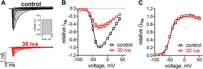
Background and purpose: Ivabradine is clinically administered to lower the heart rate, proposedly by inhibiting hyperpolarization-activated cyclic nucleotide-gated cation channels in the sinoatrial node. Recent evidence suggests that voltage-gated sodium channels (VGSC) are inhibited within the same concentration range. VGSCs are expressed within the sinoatrial node and throughout the conduction system of the heart. A block of these channels thus likely contributes to the established and newly raised clinical indications of ivabradine. We, therefore, investigated the pharmacological action of ivabradine on VGSCs in sufficient detail in order to gain a better understanding of the pro- and anti-arrhythmic effects associated with the administration of this drug. Experimental Approach: Ivabradine was tested on VGSCs in native cardiomyocytes isolated from mouse ventricles and the His-Purkinje system and on human Nav1.5 in a heterologous expression system. We investigated the mechanism of channel inhibition by determining its voltage-, frequency-, state-, and temperature-dependence, complemented by a molecular drug docking to the recent Nav1.5 cryoEM structure. Automated patch-clamp experiments were used to investigate ivabradine-mediated changes in Nav1.5 inactivation parameters and inhibition of different VGSC isoforms. Key results: Ivabradine inhibited VGSCs in a voltage- and frequency-dependent manner, but did not alter voltage-dependence of activation and fast inactivation, nor recovery from fast inactivation. Cardiac (Nav1.5), neuronal (Nav1.2), and skeletal muscle (Nav1.4) VGSC isoforms were inhibited by ivabradine within the same concentration range, as were sodium currents in native cardiomyocytes isolated from the ventricles and the His-Purkinje system. Molecular drug docking suggested an interaction of ivabradine with the classical local anesthetic binding site. Conclusion and Implications: Ivabradine acts as an atypical inhibitor of VGSCs. Inhibition of VGSCs likely contributes to the heart rate lowering effect of ivabradine, in particular at higher stimulation frequencies and depolarized membrane potentials, and to the observed slowing of intra-cardiac conduction. Inhibition of VGSCs in native cardiomyocytes and across channel isoforms may provide a potential basis for the anti-arrhythmic potential as observed upon administration of ivabradine.

Keywords: S16257; atypical inhibitor; conduction cell; ivabradine; ventricular cardiomyocyte; voltage-gated sodium channel.


Conflict of interest statement

The authors declare that the research was conducted in the absence of any commercial or financial relationships that could be construed as a potential conflict of interest.

Figures

FIGURE 1
FIGURE 1
Effect of ivabradine on the voltage-dependence on activation in human Nav1.5 channels. tsA-201 cells expressing human Nav1.5 channels were held at a holding potential of −100 mV and sodium currents were elicited by 25 ms, rectangular steps to various voltages (in 10 mV increment, see inset in A). (A) Original current traces as elicited by the voltage-clamp protocol in the absence (top, black) and presence of 30 µM ivabradine (bottom, red). (B) Current-voltage relationships as derived from the maximal inward current at the applied voltages. (C) Conductance-voltage relationships as derived from B. No significant differences were found between the half point of voltage-dependent activation, V1/2 = −48 ± 1 mV and −48 ± 1 mV (p = 0.7), and the steepness of activation, k = 9.7 ± 0.9 and 8.1 ± 0.6 (p = 0.15), for control (n = 5) and in the presence of ivabradine (n = 9), respectively.
FIGURE 2
FIGURE 2
Effect of ivabradine on the steady-state fast inactivation in human Nav1.5 channels. (A) Voltage-clamp protocol and original current traces in the presence and absence of 30 µM ivabradine. tsA-201 cells expressing human Nav1.5 channels were held at a holding potential of −140 mV. An inactivating prepulse of 50 ms to various voltages preceded a 25 ms test pulse to −10 mV, to maximally activate the available channels (inset). Current traces for a prepulse to −90 mV are highlighted in a darker color. (B) Voltage-dependence of steady-state fast inactivation as derived from maximal inward current amplitude plotted against prepulse voltages. No significant difference was found between the half point, V1/2 = −90 ± 3 and −88 ± 2 (p = 0.6), and the steepness, k = 7.8 ± 0.3 and 8.7 ± 0.4 (p = 0.12), under control conditions (n = 6) and in the presence of ivabradine (n = 7), respectively.
FIGURE 3
FIGURE 3
Automated patch-clamp analysis of the effect of ivabradine on human Nav1.5 fast inactivation. (A) Voltage-clamp protocol to simultaneously test for recovery from inactivation (RFI, blue bracket) and voltage-dependence of steady-state inactivation (SSI, red bracket) continuously once per second throughout the experiment. (B) Top: sodium current amplitude from hNav1.5 channels stably expressed in HEK-293 cells. Mean current trace ± SD from n = 11 recording channels, with each channel recording the average of a maximum of 20 cells. Perfusion of different drugs is indicated by the above bars; 100 µM riluzole (100 ril), 300 µM lidocaine (300 lid), and increasing concentrations of ivabradine (1–100 μM; 1-100 iva). Middle: the parameters from automated fitting of the SSI plot at 1 s time resolution. Half point of voltage-dependent inactivation (V1/2; left axis, red) and steepness of respective voltage-dependence (k; right axis, blue). Bottom: Selected parameters from automated fitting of the RFI plot. Recovery was fit with a double-exponential function with the second, slow time constant constrained to 150 ms (see Methods). Relative amplitude of the fast time constant of recovery (A1; left axis, red) and respective fast time constant (τ1; right axis, blue). Time axis applies to all panels in B.
FIGURE 4
FIGURE 4
Voltage-dependence of Nav1.5 inhibition by ivabradine. (A) tsA-201 cells expressing human Nav1.5 channels were held at different holding potentials (Vh = −160 to −100 mV) and depolarized to -10 mV for 25 ms every second. After 60 s, 30 µM ivabradine was applied for 3 min (red bar). Current inhibition was fitted with a mono-exponential function to derive steady-state inhibition levels. Measurements at Vh = −100 mV were corrected for Nav1.5 current rundown caused by the mentioned time-dependent shift of inactivation in whole-cell experiments. (B) Summary of Nav1.5 current amplitude in the presence of 30 µM normalized to respective control before drug application and plotted over the applied Vh. Data for Vh of −160, −140, −120, −100 mV are from n = 5, 5, 5, 5 independent measurements. (C) Summary of time constants as derived from single exponential fits to the wash-in of 30 µM ivabradine in (A). All the values are given as mean ± SEM.
FIGURE 5
FIGURE 5
Frequency dependence of Nav1.5 inhibition by ivabradine. tsA-201 cells expressing human Nav1.5 channels were held at a holding potential of −100 mV and depolarized to -10 mV for 25 ms at a frequency of either 0.1, 1, or 10 Hz. (A) Maximal peak inward sodium current amplitude during −10 mV depolarizations was monitored over time. After stable amplitude had been established 30 µM of ivabradine was washed in. Current amplitude values in the continuous presence of 30 µM ivabradine were normalized to respective control values before drug application (norm Nav1.5 current). The level of inhibition was significantly lower at 10 Hz compared to 0.1 and 1 Hz pulse frequency. (B) Summary of inhibition levels as derived from single-exponential fits to the data shown in A; mean ± SEM from n = 5, 5, and 5 independent experiments for 0.1, 1, and 10 Hz, respectively.
FIGURE 6
FIGURE 6
Temperature- and use-dependence of hNav1.5 inhibition by ivabradine. tsA-201 cells expressing human Nav1.5 channels were held at a holding potential of −100 mV and sodium currents were elicited by depolarizing voltage steps to −10 mV for 250 ms every second. Normalized maximal Nav1.5 current amplitude in the absence (first 60 s) and presence of 30 µM ivabradine (3 min application, as indicated) at a temperature of either 22°C (A) or 37°C (B). (C) Nav1.5 current inhibition at different depolarization times and temperatures. Values for depolarization times of 25 and 250 ms are plotted in the 1st and 2nd column, respectively (values for 25 ms depolarization are replotted from Figure 5). Values for temperatures of 22 and 37°C with depolarization for 250 ms are plotted in the 2nd and 3rd columns, respectively. (D) As in C, but for the wash-in time constants. Data are expressed as mean ± SEM (n = 5, 8, 5).
FIGURE 7
FIGURE 7
Concentration-dependent inhibition of different VGSC isoforms. Automated patch-clamp recordings of sodium currents through Nav1.2, Nav1.4, and Nav1.5 channels. Sodium currents were elicited from a holding potential of −150 mV by 25 ms depolarizing voltage steps to −10 mV. Ivabradine was applied at increasing concentrations (1, 3, 10, 30, 100 µM) for 3 min each. Mean data from 12, 15, and 12 channels are shown for Nav1.2, Nav1.4, and Nav1.5, respectively. Data were fit to a Hill equation with a Hill coefficient of nH = 1 to estimate half points of inhibition. IC50 values (mean ± SEM) amounted to 296 ± 11, 257 ± 15, and 137 ± 8 µM for Nav1.2, Nav1.4, and Nav1.5. One-way ANOVA with Tukey’s post-hoc test revealed a statistical difference between Nav1.5 and Nav1.2 (p < 0.001) and Nav1.4 (p < 0.001), but not between Nav1.2 and Nav1.4.
FIGURE 8
FIGURE 8
Effect of ivabradine on VGSCs in primary cardiomyocytes isolated from the ventricles and the conduction system of the mouse heart. (A) Transmitted light (TM) image of a typical ventricular cardiomyocyte (vCM) and a cardiomyocyte isolated from the conduction system of the heart (Connexin40-eGFP positive Purkinje fiber (PF); the green fluorescent light channel is overlaid with TM channel). (B) Tyoical original sodium current traces in a ventricular cardiomyocyte elicited from a holding potential of -70 mV by various voltage steps between −60 and +20 mV. Sodium currents under control conditions and in the presence of 30 µM ivabradine (30 iva; in red). (C) As in B but for Connexin40-eGFP positive cardiomyocytes isolated from the cardiac conduction system. (D) Summary of fractional sodium current levels before (control) and after equilibration with 30 µM ivabradine derived from n = 7 ventricular cardiomyocytes. (E) Summary of fractional sodium current levels before (control) and after equilibration with 30 µM ivabradine derived from n = 7 Purkinje fibers. Data are given as mean ± SEM.
FIGURE 9
FIGURE 9
Molecular interactions of ivabradine with Nav1.5. (A) Chemical structure of ivabradine as used for the drug docking with a charged amine nitrogen at physiological pH. (B) Representative docking pose of Ivabradine shown as purple spheres in the cavity of Nav1.5 (cartoon representation) viewed from the top. (C) Side view of the binding site below the selectivity filter, with the voltage sensor of domain II hidden for clarity. (D) Residues within 5 Å of ivabradine are shown in stick representation. Dotted lines between F934 and Ivabradine denote π- π interactions. The distance between the two benzene rings is 4.1 Å.

Similar articles

Cited by

References

    1. Abraham S., Beatch G. N., MacLeod B. A., Walker M. J. (1989). Antiarrhythmic Properties of Tetrodotoxin against Occlusion-Induced Arrhythmias in the Rat: A Novel Approach to the Study of the Antiarrhythmic Effects of Ventricular Sodium Channel Blockade. J. Pharmacol. Exp. Ther. 251, 1166–1173. - PubMed
    1. Al-Ghamdi S., Al-Fayyadh M. I., Hamilton R. M. (2013). Potential New Indication for Ivabradine: Treatment of a Patient with Congenital Junctional Ectopic Tachycardia. J. Cardiovasc. Electrophysiol. 24, 822–824. 10.1111/jce.12081 - DOI - PubMed
    1. Amstetter D., Badt F., Rubi L., Bittner R. E., Ebner J., Uhrin P., et al. (2021). The Bradycardic Agent Ivabradine Decreases Conduction Velocity in the AV Node and in the Ventricles In-Vivo . Eur. J. Pharmacol. 893, 173818. 10.1016/j.ejphar.2020.173818 - DOI - PubMed
    1. Baruscotti M., DiFrancesco D., Robinson R. B. (1996). A TTX-Sensitive Inward Sodium Current Contributes to Spontaneous Activity in Newborn Rabbit Sino-Atrial Node Cells. J. Physiol. 492, 21–30. 10.1113/jphysiol.1996.sp021285 - DOI - PMC - PubMed
    1. Bean B. P., Cohen C. J., Tsien R. W. (1983). Lidocaine Block of Cardiac Sodium Channels. J. Gen. Physiol. 81, 613–642. 10.1085/jgp.81.5.613 - DOI - PMC - PubMed

LinkOut - more resources