. 2022 May 9:2022:2292481.
doi: 10.1155/2022/2292481. eCollection 2022.

DNAJA1 Stabilizes EF1A1 to Promote Cell Proliferation and Metastasis of Liver Cancer Mediated by miR-205-5p

Affiliations

DNAJA1 Stabilizes EF1A1 to Promote Cell Proliferation and Metastasis of Liver Cancer Mediated by miR-205-5p

Lizhi Yi et al. J Oncol. .

Abstract

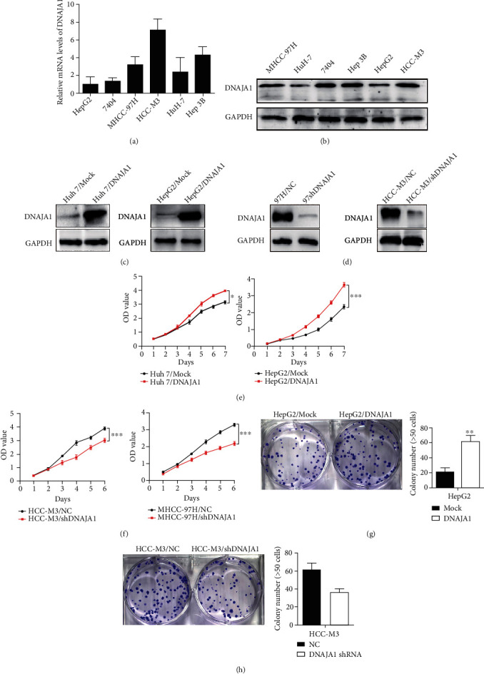
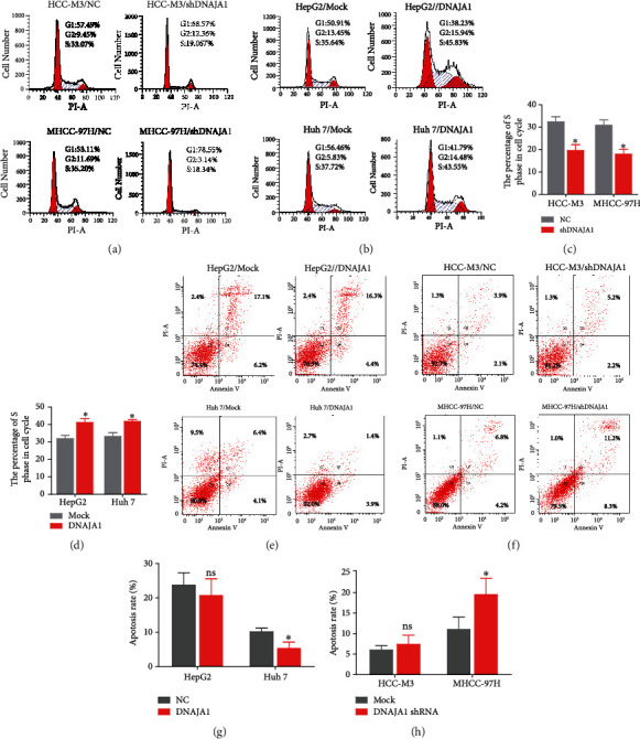
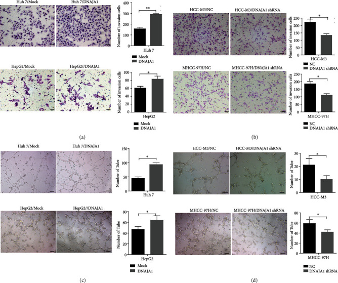
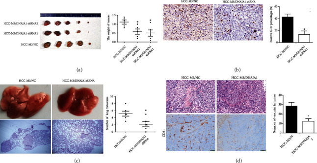
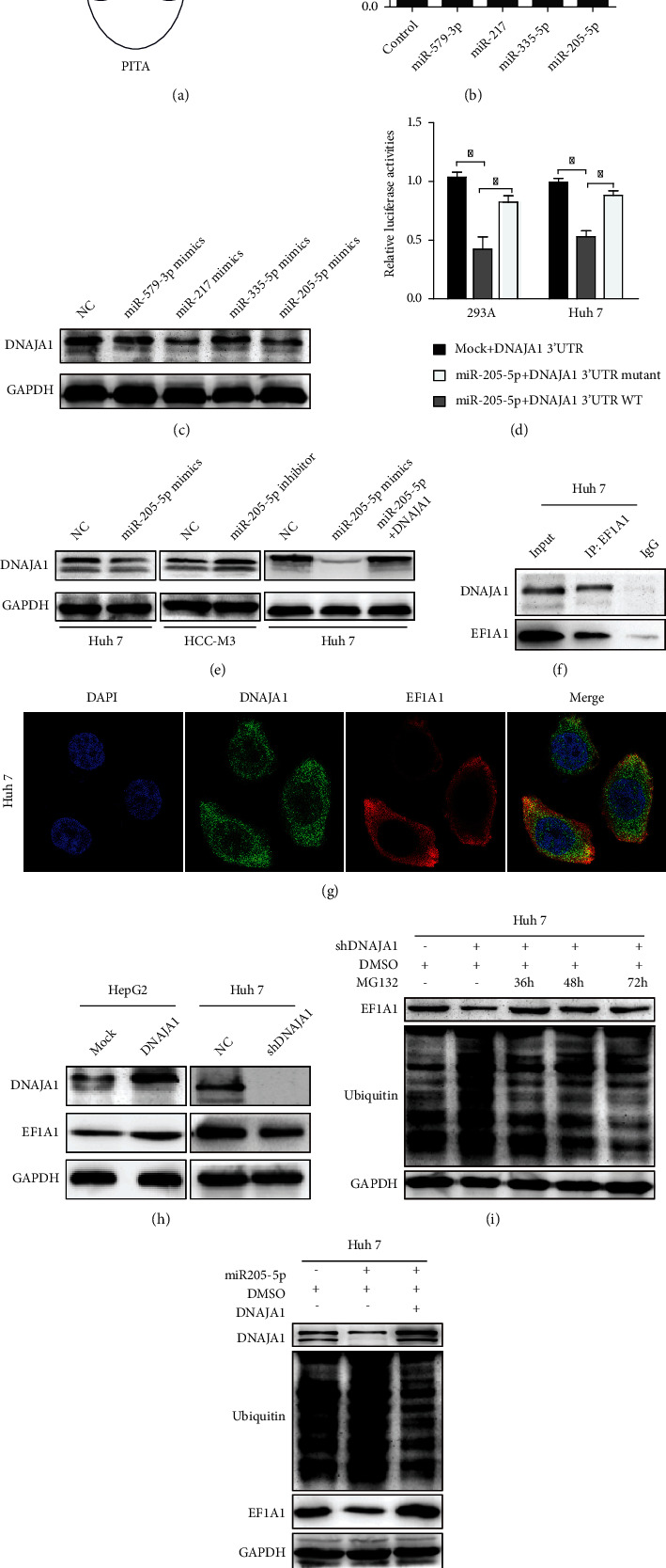
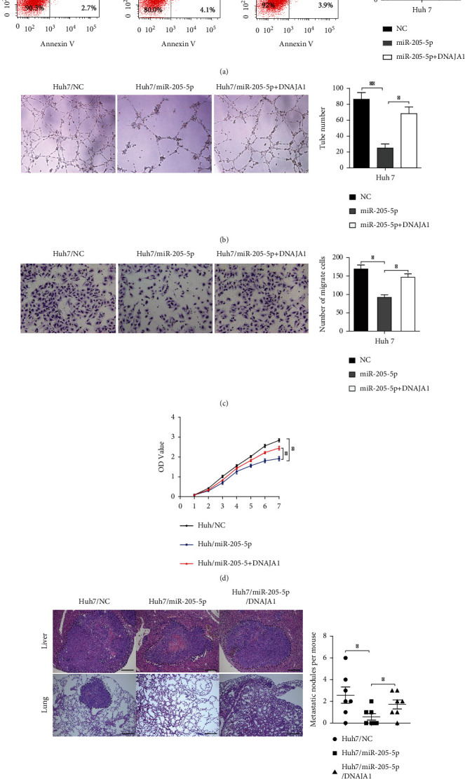
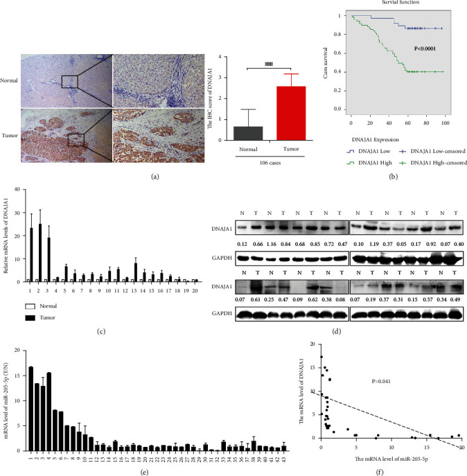
Liver cancer is one of the most common and aggressive malignancies worldwide with poor prognosis. Studies on pathogenesis of liver cancer are urgently demanded to develop better treatment strategy. Here, we found that overexpression of DnaJ heat shock protein family (Hsp40) member A1 (DNAJA1) increased cell proliferation, invasion, and angiogenesis in Huh 7 and HepG2 cells, while depletion of DNAJA1 in MHCC-97H and HCC-M3 showed opposite effects. In vivo functional assays indicated that DNAJA1 promoted tumor growth and pulmonary metastasis in mice. Mechanistically, as a direct target of miR-205-5p, DNAJA1 promoted proliferation and metastasis of liver cancer cells by stabilizing eukaryotic elongation factor 1A1 (EF1A1). Moreover, DNAJA was markedly upregulated in liver cancer tissues (P < 0.05) and was significantly associated with poor prognosis. And its expression was correlated with differentiation (P < 0.001), dissemination (P < 0.001), and serum AFP (P = 0.029). The mRNA levels of miR-205-5p and DNAJA1 were negatively correlated in liver cancer. In conclusion, our study reveals that DNAJA1 acts as an oncogene in liver cancer via miR-205-5p/EF1A1 axis and might be a potential biomarker to predict the prognosis for liver cancer patients.


Conflict of interest statement

No conflicts of interest, financial or otherwise, are declared by the authors.

Figures

Figure 1
Figure 1
DNAJA1 promoted proliferation of liver cancer cells in vitro. (a) The mRNA level of DNAJA1 in six liver cancer cell lines detected by q-PCR. (b) The protein expression of DNAJA1 in six liver cancer cell lines detected by western blot. Error bars represent mean ± SD from three independent experiments. P < 0.05. (c) Relative protein expressions of DNAJA1 in Huh 7 and HepG2 cells stably transfected with DNAJA1 overexpression vector or control vector by western blot. (d) Relative protein expressions of DNAJA1 in MHCC-97H and HCC-M3 cells stably transfected with DNAJA1 shRNA or negative control by western blot. (e and f) Effect of DNAJA1 overexpression or downregulation on cell proliferation of Huh 7 and HepG2 cells or MHCC-97H and HCC-M3 cells detected by CCK-8 assay. (g and h) Effect of DNAJA1 overexpression or inhibition on cell clone formation ability of HepG2 cells or HCC-M3 cells.
Figure 2
Figure 2
DNAJA1 promotes the proliferation by arresting the tumor cells at the S and G2/M phase and reducing apoptosis. (a) Representative histograms depicting cell cycle profiles of MHCC-97H and HCC-M3 cells stably transfected with DNAJA1 shRNA or negative control. (b) Representative histograms depicting cell cycle profiles of Huh 7 and HepG2 cells stably transfected with DNAJA1 overexpression vector or control vector. (c and d) The quantification of the effect of DNAJA1 on cell cycle. (e) Percent of apoptosis in Huh 7 and HepG2 cells stably transfected with DNAJA1 overexpression vector or control vector. (f) Percent of apoptosis in MHCC-97H and HCC-M3 cells stably transfected with DNAJA1 shRNA or negative control. (g and h) The quantification of the effect of DNAJA1 on apoptosis. Error bars represent mean ± SD from three independent experiments. P < 0.05.
Figure 3
Figure 3
DNAJA1 promoted invasion and angiogenesis of liver cancer cells in vitro. (a) The effect of DNAJA1 on invasion in Huh 7 and HepG2 cells stably transfected with DNAJA1 overexpression vector or control vector. (b) The effect of DNAJA1 on invasion in MHCC-97H and HCC-M3 cells stably transfected with DNAJA1 shRNA or negative control. (c) The effect of DNAJA1 on angiogenesis in Huh 7 and HepG2 cells stably transfected with DNAJA1 overexpression vector or control vector. (d) The effect of DNAJA1 on angiogenesis in MHCC-97H and HCC-M3 cells stably transfected with DNAJA1 shRNA or negative control.
Figure 4
Figure 4
DNAJA1 promoted proliferation and metastasis of liver cancer cells in vivo. (a) Images and statistical analysis of subcutaneous tumor formed by HCC-M3 cells stably transfected with DNAJA1 shRNA or negative control. Qualification of tumor weight in HCC-M3/DNAJA1 shRNA or HCC-M3/NC groups. (b) Representative images of Ki67 staining in subcutaneous tumor sections by immunohistochemistry. (c) The effect of DNAJA1 overexpression on metastasis in HCC-M3/DNAJA1 shRNA or HCC-M3/NC groups. (d) The effect of DNAJA1 overexpression on angiogenesis in HCC-M3/DNAJA1 shRNA or HCC-M3/NC groups. Error bars represent mean ± SD from three independent experiments. P < 0.05.
Figure 5
Figure 5
DNAJA1 was directly targeted by miR-205-5p and interacted with EF1A1. (a) Predicted microRNAs which targeted to DNAJA1 were predicted by three common bioinformatic algorithms. (b) Luciferase assay analyses of the indicated cells transfected with the indicated reporters with miR-205-5p, miR335-5p, miR579-3p, and miR-217. (c) Western blot analysis of DNAJA1 in HCC-M3 cells transfected with miR-205-5p, miR335-5p, miR579-3p, and miR-217 mimics. (d) Luciferase assay analyses of HCC-M3 cells simultaneously transfected with the 3′-UTRs of WT and mutant DNAJA1 and miR-205-5p mimics. (e) Western blot analysis of DNAJA1 in HCC-M3 cells transfected with miR-205-5p with or without DNAJA1. (f) Mutual combination between DNAJA1 and EF1A1 in Huh 7 cells by Co-IP assay. (g) Colocalization of DNAJA1 and EF1A1 by immunofluorescence (blue: DAPI, green: DNAJA1, and red: EF1A1). (h) The protein level of DNAJA1 and EF1A1 in DNAJA1 overexpressing or inhibiting cells by western blot. (i) Effect of time-course inhibition of DNAJA1 on the expression of EF1A1 and the status of its ubiquitination by western blot. (j) Effect of miR-205-5p on the expression of EF1A1 and DNAJA1 and ubiquitin in HCC-M3 cells by western blot. GAPDH was used as internal control.
Figure 6
Figure 6
DNAJA1 reversed the effect of miR-205-5p on the liver cells. (a) Representative histograms depicting cell apoptosis of HCC-M3 cells stably transfected with miR-205-5p with or without DNAJA1 shRNA. (b) Tubule formation assays of HCC-M3 cells stably transfected with miR-205-5p with or without DNAJA1 shRNA. (c) Transwell assays of HCC-M3 cells stably transfected with miR-205-5p with or without DNAJA1 shRNA. (d) CCK8 assays of HCC-M3 cells stably transfected with miR-205-5p with or without DNAJA1 shRNA. (e) Images and statistical analysis of liver subcapsular tumor implantation of Huh 7 cells stably transfected with miR-205-5p or miR-205-5p/DNAJA1 shRNA or negative control.
Figure 7
Figure 7
DNAJA1 is upregulated in liver cancer and negatively related to miR-205-5p. (a) Representative images of DNAJA1 proteins in 106 cases of paraffin-embedded liver cancer specimens by immunohistochemistry. (b) High expression of DNAJA1 associated with shorter survival of liver cancer patients by the Kaplan-Meier analysis. (c and d) DNAJA1 mRNA and protein level in 20 cases (presented) of fresh liver cancer tissues and corresponding normal liver detected by qPCR and western blotting. (e) Detection of miR-205-5p level in 43 cases of paired fresh HCC patients by real-time PCR. (f) The relationship between miR-205-5p and DNAJA1. Error bars represent mean ± SD from three independent experiments.

Similar articles

Cited by

References

    1. Siegel R. L., Cancer M. K. J. A. Cancer statistics, 2019. CA: a Cancer Journal for Clinicians . 2019;69(1):7–34. doi: 10.3322/caac.21551. - DOI - PubMed
    1. Chen W., Zheng R., Baade P. D., et al. Cancer statistics in China, 2015. CA: a Cancer Journal for Clinicians . 2016;66(2):115–132. doi: 10.3322/caac.21338. - DOI - PubMed
    1. Bosch F. X., Ribes J., Díaz M., Cléries R. Gastroenterology (New York, N.Y. 1943) Vol. 127. United States: Elsevier BV; 2004. Primary liver cancer: eorldwide incidence and trends; pp. S5–S16. - PubMed
    1. Waly R. S., Yangde Z., Yuxiang C. Hepatocellular carcinoma: focus on different aspects of management. Oncologia . 2012;2012, article 421673:1–12. doi: 10.5402/2012/421673. - DOI - PMC - PubMed
    1. Villanueva A. Hepatocellular carcinoma. The New England Journal of Medicine . 2019;380(15):1450–1462. doi: 10.1056/NEJMra1713263. - DOI - PubMed

LinkOut - more resources