Review
. 2022 May 14:13:20417314221095997.
doi: 10.1177/20417314221095997. eCollection 2022 Jan-Dec.

Modeling the blood-brain barrier for treatment of central nervous system (CNS) diseases

Affiliations
Review

Modeling the blood-brain barrier for treatment of central nervous system (CNS) diseases

Olivia Rice et al. J Tissue Eng. .

Abstract

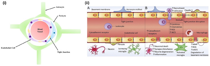
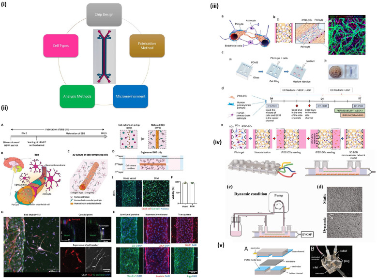
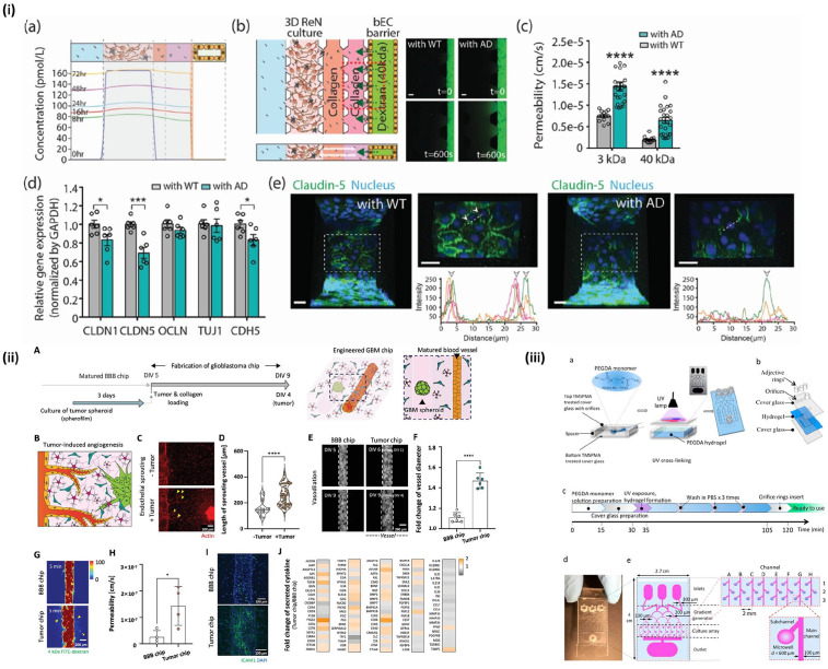
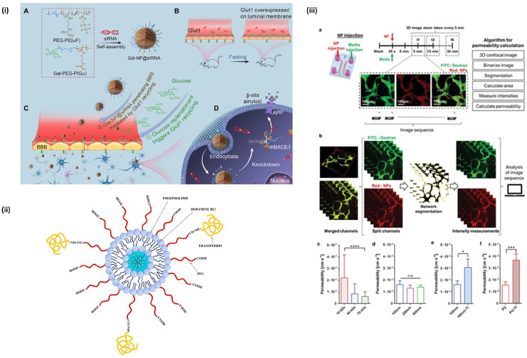
The blood-brain barrier (BBB) is the most specialized biological barrier in the body. This configuration of specialized cells protects the brain from invasion of molecules and particles through formation of tight junctions. To learn more about transport to the brain, in vitro modeling of the BBB is continuously advanced. The types of models and cells selected vary with the goal of each individual study, but the same validation methods, quantification of tight junctions, and permeability assays are often used. With Transwells and microfluidic devices, more information regarding formation of the BBB has been observed. Disease models have been developed to examine the effects on BBB integrity. The goal of modeling is not only to understand normal BBB physiology, but also to create treatments for diseases. This review will highlight several recent studies to show the diversity in model selection and the many applications of BBB models in in vitro research.

Keywords: Blood-brain-barrier; CNS; brain-on-chip; drug delivery; neurodegenerative disease.


Conflict of interest statement

Declaration of conflicting interests: The author(s) declared the following potential conflicts of interest with respect to the research, authorship, and/or publication of this article: Dr. Yupeng Chen is a co-founder of Eascra Biotech.

Figures

Figure 1.
Figure 1.
In vivo blood-brain barrier structure: (i) Cross sectional view. (ii) A. View of healthy BBB and B. view of BBB with neurodegenerative disease. Reproduced from Saraiva et al..
Figure 2.
Figure 2.
Validation techniques commonly used for BBB models: (i) TEER measurements from four different types of brain endothelial cells. Reproduced from Eigenmann et al., (ii) tight junction protein staining of cadherin and ZO-1. Reproduced from Zakharova et al., (iii) validation of models using three types of brain endothelial cells with tight junction protein staining and western blot, as well as verification of p-gp expression. Reproduced from Gericke et al.
Figure 3.
Figure 3.
Timeline visualization of the advancements in BBB modeling.
Figure 4.
Figure 4.
Transwell model for BBB studies: (i) A. Transwell monoculture with human brain endothelial cells (HBMECs). B. Transwell co-culture with HBMECs and pericytes. C. Transwell tri-culture with HBMEC, pericytes, and astrocytes. D. Transwell co-culture with HBMECs and astrocytes, (ii) fabrication of Transwell model with topographical changes for improved BBB integrity. Reproduced from Zakharova et al., (iii) four human brain endothelial cell lines cultured for BBB model optimization. Reproduced from Eigenmann et al.
Figure 5.
Figure 5.
Microfluidic devices used for BBB modeling: (i) example of a two channel microfluidic device and components of tissue models, (ii) 3D culture on chip with various validation methods. Reproduced from Seo et al., (iii) microfluidic model using a combination of PDMS, glass, and fibrin gel with a mixture of three cell types. Reproduced from Campisi et al., (iv) microfluidic chip using a pump for dynamic flow of media and built-in TEER analysis. Reproduced from Santa-Maria et al., (v) model demonstrating ability for continuous TEER measurement. Reproduced from Tu et al.
Figure 6.
Figure 6.
Studies utilizing disease models of BBB: (i) comparison of healthy (WT) BBB with Alzheimer’s disease (AD) BBB in permeability and expression of tight junction proteins. Reproduced from Shin et al., (ii, iii) BBB models utilizing tumor spheroids within the tissue chip. Reproduced from Seo et al. and Fan et al.
Figure 7.
Figure 7.
Nanoparticles for BBB drug delivery: (i) example of a NP used for RNAi therapy in AD treatment. Reproduced from Zhou et al., (ii) a lipid nanoparticle with transferrin ligand for brain endothelial cell targeting. Reproduced from Lopalco et al., (iii) study using machine learning for optimization of NP delivery. Reproduced from Lee et al.

Similar articles

Cited by

References

    1. Pandit R, Chen L, Götz J. The blood-brain barrier: physiology and strategies for drug delivery. Adv Drug Deliv Rev 2020; 165–166:1–14. - PubMed
    1. Saraiva C, Praça C, Ferreira R, et al.. Nanoparticle-mediated brain drug delivery: overcoming blood-brain barrier to treat neurodegenerative diseases. J Control Release 2016; 235:34–47. - PubMed
    1. Abbott NJ, Patabendige AA, Dolman DE, et al.. Structure and function of the blood-brain barrier. Neurobiol Dis 2010; 37(1): 13–25. - PubMed
    1. Helms HC, Abbott NJ, Burek M, et al.. In vitro models of the blood-brain barrier: an overview of commonly used brain endothelial cell culture models and guidelines for their use. J Cereb Blood Flow Metab 2016; 36(5): 862–890. - PMC - PubMed
    1. Bernacki J, Dobrowolska A, Nierwińska K, et al.. Physiology and pharmacological role of the blood-brain barrier. Pharmacol Rep 2008; 60(5): 600–622. - PubMed

LinkOut - more resources