

In Vivo Characterization of a Red Light-Activated Vasodilation: A Photobiomodulation Study
- PMID: 35586710
- PMCID: PMC9108481
- DOI: 10.3389/fphys.2022.880158
In Vivo Characterization of a Red Light-Activated Vasodilation: A Photobiomodulation Study
Abstract
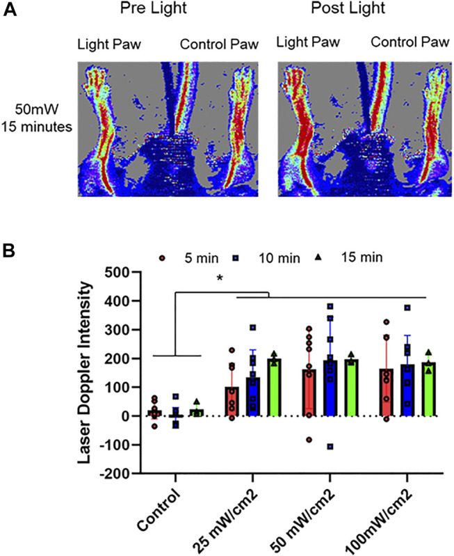
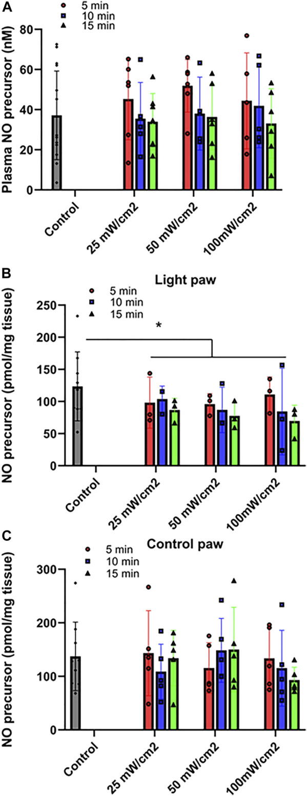
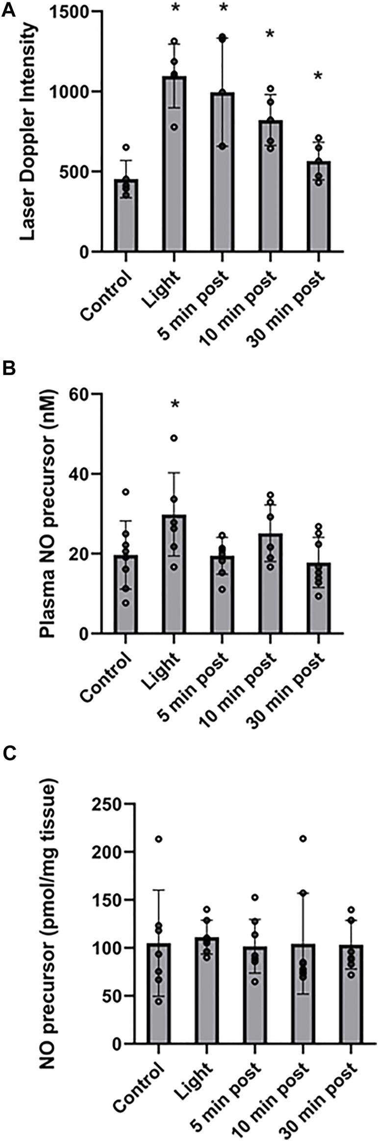
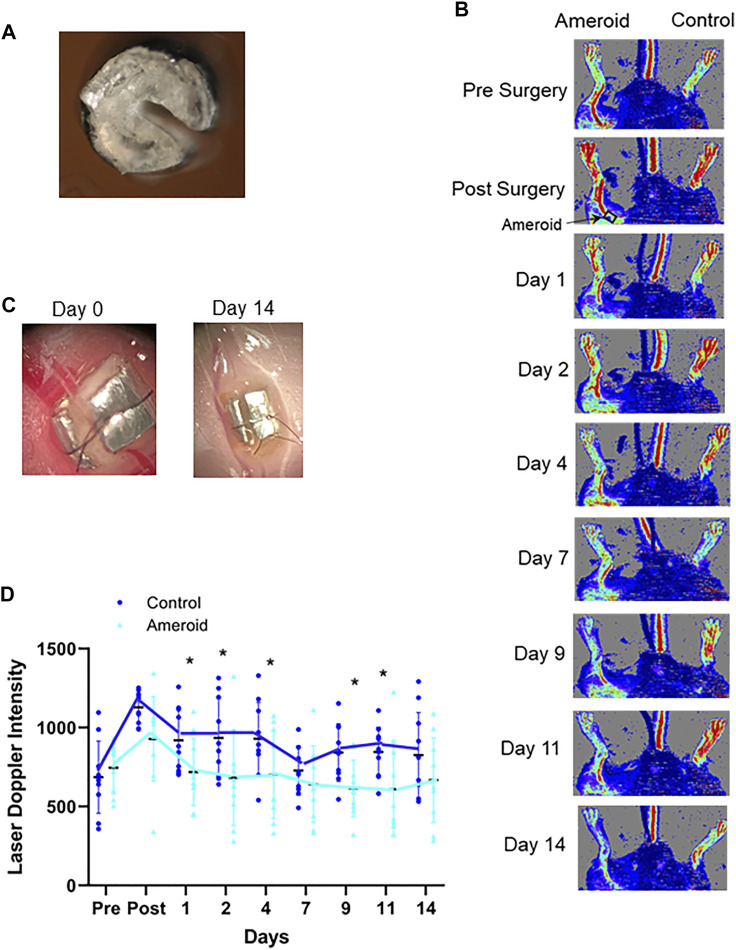
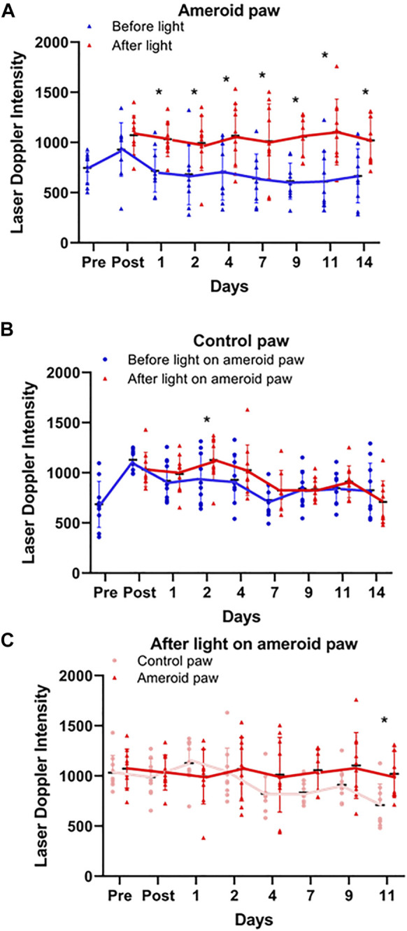
Nitric oxide dependent vasodilation is an effective mechanism for restoring blood flow to ischemic tissues. Previously, we established an ex vivo murine model whereby red light (670 nm) facilitates vasodilation via an endothelium derived vasoactive species which contains a functional group that can be reduced to nitric oxide. In the present study we investigated this vasodilator in vivo by measuring blood flow with Laser Doppler Perfusion imaging in mice. The vasodilatory nitric oxide precursor was analyzed in plasma and muscle with triiodide-dependent chemiluminescence. First, a 5-10 min irradiation of a 3 cm2 area in the hind limb at 670 nm (50 mW/cm2) produced optimal vasodilation. The nitric oxide precursor in the irradiated quadriceps tissue decreased significantly from 123 ± 18 pmol/g tissue by both intensity and duration of light treatment to an average of 90 ± 17 pmol/g tissue, while stayed steady (137 ± 21 pmol/g tissue) in unexposed control hindlimb. Second, the blood flow remained elevated 30 min after termination of the light exposure. The nitric oxide precursor content significantly increased by 50% by irradiation then depleted in plasma, while remained stable in the hindlimb muscle. Third, to mimic human peripheral artery disease, an ameroid constrictor was inserted on the proximal femoral artery of mice and caused a significant reduction of flow. Repeated light treatment for 14 days achieved steady and significant increase of perfusion in the constricted limb. Our results strongly support 670 nm light can regulate dilation of conduit vessel by releasing a vasoactive nitric oxide precursor species and may offer a simple home-based therapy in the future to individuals with impaired blood flow in the leg.
Keywords: endothelium; nitric oxide; photobiomodulation; red light therapy; vasodilation.
Copyright © 2022 Keszler, Lindemer, Broeckel, Weihrauch, Gao and Lohr.
Conflict of interest statement
The authors declare that the research was conducted in the absence of any commercial or financial relationships that could be construed as a potential conflict of interest.
Figures

Similar articles
-
Red/near infrared light stimulates release of an endothelium dependent vasodilator and rescues vascular dysfunction in a diabetes model.Free Radic Biol Med. 2017 Dec;113:157-164. doi: 10.1016/j.freeradbiomed.2017.09.012. Epub 2017 Sep 19. Free Radic Biol Med. 2017. PMID: 28935419 Free PMC article.
-
Assessment of tissue perfusion and vascular function in mice by scanning laser Doppler perfusion imaging.Biochem Pharmacol. 2020 Jun;176:113893. doi: 10.1016/j.bcp.2020.113893. Epub 2020 Mar 3. Biochem Pharmacol. 2020. PMID: 32135157
-
Red light mediates the exocytosis of vasodilatory vesicles from cultured endothelial cells: a cellular, and ex vivo murine model.Photochem Photobiol Sci. 2024 Feb;23(2):355-364. doi: 10.1007/s43630-023-00522-1. Epub 2024 Jan 26. Photochem Photobiol Sci. 2024. PMID: 38277065 Free PMC article.
-
Red light stimulates vasodilation through extracellular vesicle trafficking.J Photochem Photobiol B. 2021 Jul;220:112212. doi: 10.1016/j.jphotobiol.2021.112212. Epub 2021 May 12. J Photochem Photobiol B. 2021. PMID: 34049180 Free PMC article.
-
Control of skeletal muscle blood flow during dynamic exercise: contribution of endothelium-derived nitric oxide.Sports Med. 1996 Feb;21(2):119-46. doi: 10.2165/00007256-199621020-00004. Sports Med. 1996. PMID: 8775517 Review.
Cited by
-
Morphological features of the photoplethysmographic signal: a new approach to characterize the microcirculatory response to photobiomodulation.Front Physiol. 2023 Sep 25;14:1175470. doi: 10.3389/fphys.2023.1175470. eCollection 2023. Front Physiol. 2023. PMID: 37817983 Free PMC article.
-
Assessment of photobiomodulation in response to the microcirculation in arteriovenous fistula for hemodialysis patient.Asian Biomed (Res Rev News). 2025 Feb 28;19(1):3-13. doi: 10.2478/abm-2025-0005. eCollection 2025 Feb. Asian Biomed (Res Rev News). 2025. PMID: 40231166 Free PMC article.
-
Vascular Responses following Light Therapy: A Pilot Study with Healthy Volunteers.J Clin Med. 2023 Mar 13;12(6):2229. doi: 10.3390/jcm12062229. J Clin Med. 2023. PMID: 36983231 Free PMC article.
-
Light buckets and laser beams: mechanisms and applications of photobiomodulation (PBM) therapy.Geroscience. 2025 Jun;47(3):2777-2789. doi: 10.1007/s11357-025-01505-z. Epub 2025 Jan 18. Geroscience. 2025. PMID: 39826026 Free PMC article. Review.
-
Effect of Photobiomodulation on Protein Kinase Cδ, Cytochrome C, and Mitochondria in U87 MG Cells.Cells. 2023 May 22;12(10):1441. doi: 10.3390/cells12101441. Cells. 2023. PMID: 37408275 Free PMC article.
References
-
- Adams M. R., McCredie R., Jessup W., Robinson J., Sullivan D., Celermajer D. S. (1997). Oral L-Arginine Improves Endothelium-Dependent Dilatation and Reduces Monocyte Adhesion to Endothelial Cells in Young Men with Coronary Artery Disease. Atherosclerosis 129, 261–269. 10.1016/s0021-9150(96)06044-3 - DOI - PubMed
-
- Amaroli A., Pasquale C., Zekiy A., Utyuzh A., Benedicenti S., Signore A., et al. (2021). Photobiomodulation and Oxidative Stress: 980 Nm Diode Laser Light Regulates Mitochondrial Activity and Reactive Oxygen Species Production. Oxid Med. Cel Longev 2021, 6626286. 10.1155/2021/6626286 - DOI - PMC - PubMed
Grants and funding
LinkOut - more resources
Full Text Sources