

The function of the tensor tympani muscle: a comprehensive review of the literature
- PMID: 35586903
- PMCID: PMC9256479
- DOI: 10.5115/acb.21.032
The function of the tensor tympani muscle: a comprehensive review of the literature
Abstract
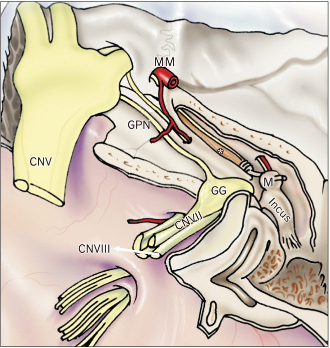
The tensor tympani muscle is structurally important in the middle ear, specifically through its involvement in the impedance of sound in response to intense auditory and non-auditory stimuli. Despite numerous studies, its true function has been debated for many years; questions still remain about its role in auditory and non-auditory reflexes and in sound damping. Some studies suggest that the tensor tympani muscle contracts as a result of non-auditory stimulation such as facial or head movements; others suggest that it contracts due to input from the cochlear nucleus, therefore by way of auditory stimulation. Whatever the cause, contraction of the tensor tympani muscle results in low frequency mixed hearing loss, either to protect the inner ear from loud sounds or to desensitize the ear to self-generated sounds. A review of these studies indicated that the tensor tympani muscle has a wide range of functions, yet the mechanisms of some of them have not been clearly demonstrated. One major question is whether the tensor tympani muscle contributes to sound damping; and if it does, what specific role it serves. The primary purpose of this review article is to explore the functions of the tensor tympani muscle in light of recent research advances.
Keywords: Anatomy; Ear; Hearing; Muscles; Sound.
Conflict of interest statement
No potential conflict of interest relevant to this article was reported.
Figures
References
-
- Møller AR. Hearing: anatomy, physiology, and disorders of the auditory system. 2nd ed. Elsevier; Amsterdam: 2006.
-
- Standring S. Gray's anatomy: the anatomical basis of clinical practice. 41st ed. Elsevier; London: 2015.
Publication types
LinkOut - more resources
Full Text Sources