. 2022 Dec;76(6):1794-1810.
doi: 10.1002/hep.32577. Epub 2022 Jun 30.

Hepatocellular cystathionine γ lyase/hydrogen sulfide attenuates nonalcoholic fatty liver disease by activating farnesoid X receptor

Affiliations

Hepatocellular cystathionine γ lyase/hydrogen sulfide attenuates nonalcoholic fatty liver disease by activating farnesoid X receptor

Wenjing Xu et al. Hepatology. 2022 Dec.

Abstract

Background and aims: Hydrogen sulfide (H2 S) plays a protective role in NAFLD. However, whether cystathionine γ lyase (CSE), a dominant H2 S generating enzyme in hepatocytes, has a role in the pathogenesis of NAFLD is currently unclear.

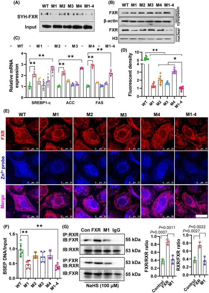
Approach and results: We showed that CSE protein expression is dramatically downregulated, especially in fibrotic areas, in livers from patients with NAFLD. In high-fat diet (HFD)-induced NAFLD mice or an oleic acid-induced hepatocyte model, the CSE/H2 S pathway is also downregulated. To illustrate a regulatory role for CSE in NAFLD, we generated a hepatocyte-specific CSE knockout mouse (CSELKO ). Feeding an HFD to CSELKO mice, they showed more hepatic lipid deposition with increased activity of the fatty acid de novo synthesis pathway, increased hepatic insulin resistance, and higher hepatic gluconeogenic ability compared to CSELoxp control mice. By contrast, H2 S donor treatment attenuated these phenotypes. Furthermore, the protection conferred by H2 S was blocked by farnesoid X receptor (FXR) knockdown. Consistently, serum deoxycholic acid and lithocholic acid (FXR antagonists) were increased, and tauro-β-muricholic acid (FXR activation elevated) was reduced in CSELKO . CSE/H2 S promoted a post-translation modification (sulfhydration) of FXR at Cys138/141 sites, thereby enhancing its activity to modulate expression of target genes related to lipid and glucose metabolism, inflammation, and fibrosis. Sulfhydration proteomics in patients' livers supported the CSE/H2 S modulation noted in the CSELKO mice.

Conclusions: FXR sulfhydration is a post-translational modification affected by hepatic endogenous CSE/H2 S that may promote FXR activity and attenuate NAFLD. Hepatic CSE deficiency promotes development of nonalcoholic steatohepatitis. The interaction between H2 S and FXR may be amenable to therapeutic drug treatment in NAFLD.


Conflict of interest statement

Nothing to report.

Figures

FIGURE 1
FIGURE 1
Downregulation of hepatic cystathionine γ lyase (CSE)/hydrogen sulfide (H2S) in patients and mice with NAFLD. (A) Immunohistochemistry (IHC) staining of CSE in liver biopsy from patients with and without NAFLD. Protein expression was evaluated by an H‐score semiquantitative approach. Bar = 20 μm. Red line area is a fibrotic area. There is no positive CSE staining in this area. (B) Endogenous H2S generation key enzymes: cystathionine β synthase (CBS), CSE, and 3‐mercaptopyruvate sulfur transferase (3‐MST) messenger RNA (mRNA) expression, protein expression by western blot (C) or IHC staining, bar = 20 μm (D) in high‐fat diet (HFD)‐induced NAFLD mice. N = 5. (E) H2S generation ratio of mouse liver tissue was measured by methylene blue assay. N = 7–8. (F) Oleic acid (OA; 400 μM) treated primary mouse hepatocytes for 48 h, LipidTox staining for lipid deposition, Mito‐HS for H2S production. (G) CBS, CSE, and 3‐MST protein expression were measured in OA‐treated hepatocytes. N = 6.
FIGURE 2
FIGURE 2
Hepatic cystathionine γ lyase (CSE) deletion mice exacerbated high‐fat diet (HFD)‐induced NAFLD. (A) General pathology (bar = 1 cm) and Oil Red O staining (bar = 50 μm) for liver tissues in hepatic CSE‐specific knockout mice (CSE LKO) and control loxp/loxp mice (CSE Loxp). (B) Hematoxylin–eosin (H&E) and Sirius Red staining for liver tissues. Bar = 50 μm. (C) Liver triglyceride (TG) and total cholesterol (TC) levels in NAFLD mice. (D) Oral glucose tolerance test (OGTT), (E) insulin tolerance test (ITT), (F) and pyruvate tolerance test (PTT) were compared between CSE LKO and CSE Loxp. N = 8–10 in all animal experiments. (G) Lipid metabolism–related genes expression was assayed by quantitative real‐time PCR. N = 6. **p < 0.01. (F) Fatty acid de novo synthesis–related protein expression was detected by western blot. N = 6.
FIGURE 3
FIGURE 3
Hydrogen sulfide (H2S) donor treatment ameliorated high‐fat diet (HFD)‐induced NAFLD and glucose metabolism disorder. In HFD‐induced NAFLD mouse model, H2S donor‐NaHS or GYY4137 treatment for 12 weeks, then hematoxylin–eosin (H&E) staining (A), Oil Red O staining (B) was used for evaluation the lipid deposition in liver. Bar = 50 μm. (C) Changes of serum triglycerides (TG), total cholesterol (TC), and liver TG, TC level after H2S donor treatment. *p < 0.05; **p < 0.01. (D) Oral glucose tolerance test (OGTT), (E) insulin tolerance test (ITT), and (F) pyruvate tolerance test (PTT) changes after H2S donor treatment. Two‐way mixed effect analysis of variance (ANOVA) was used, **p < 0.01 vs. normal chow, # p < 0.05 vs. HFD. (G) Fatty acid de novo synthesis–related protein expression by western blot. (H) The semiquantitative analysis of above proteins by relative gray density plus area of target protein comparison to β‐actin. **p < 0.01. Six independent experiments were performed.
FIGURE 4
FIGURE 4
Hepatocellular endogenous cystathionine γ lyase (CSE)/hydrogen sulfide (H2S) upregulated farnesoid X receptor (FXR) expression. Total RNA was extracted from about 30 mg liver tissue of CSE LKO or CSE Loxp mice, then bulk RNA‐sequencing was performed. (A) Volcano plot shows the downregulated genes (blue) and upregulated genes (red) in CSE LKO compared with CSE Loxp mice. Open circles represent the Gene Ontology (GO)‐enriched genes. (B) GO analysis shows the changed genes enrichment in the major pathway. (C) FXR protein expression in CSE knockout liver tissues. N = 6. (D) Hepatic FXR protein level changes after H2S donor treatment. With overexpression CSE by adenovirus or knockdown CSE by siRNA in hepG2 cells, the FXR messenger RNA (mRNA) (E) and protein expression (F) change. N = 6. Then, the fatty acid de novo synthesis–related genes: sterol response element binding protein 1c (SREBP‐1c), acetyl‐CoA carboxylase (ACC), fatty acid synthase (FAS), and stearoyl‐CoA desaturase 1 (SCD1) mRNA (N = 4) (G) and protein expression (H) changes associated with CSE overexpression or knockdown. *p < 0.05; **p < 0.01. N = 6.
FIGURE 5
FIGURE 5
Knockdown farnesoid X receptor (FXR) by lentivirus blocked hydrogen sulfide (H2S) donor's protection on NAFLD. Knockdown FXR shRNA lentivirus (10 multiplicity of infection [MOI]) was bolus injected by tail vein before high‐fat diet (HFD) feeding. Continue feeding HFD for 12 weeks; hematoxylin–eosin (H&E) staining for liver tissue morphology, Oil Red O staining for hepatic lipid deposition. Bar = 100 μm (A). (B) Serum and liver triglycerides (TG), total cholesterol (TC) level were measured. *p < 0.05, **p < 0.01. (C) Oral glucose tolerance test (OGTT), (D) insulin tolerance test (ITT), and (E) pyruvate tolerance test (PTT) were assayed for glucose metabolism. *p < 0.05, **p < 0.01. N = 8 in this animal experiment. (F) Hepatic FXR protein, fatty acid de novo synthesis genes: sterol response element binding protein 1c (SREBP‐1c), acetyl‐CoA carboxylase (ACC), fatty acid synthase (FAS), and stearoyl‐CoA desaturase (SCD) protein expression was detected by western blot. N = 6. (G) The semiquantitative analysis of above proteins. *p < 0.05, **p < 0.01.
FIGURE 6
FIGURE 6
Cystathionine γ lyase (CSE)/hydrogen sulfide (H2S) sulfhydrated farnesoid X receptor (FXR) and promoted its binding to Zn2+, then enhanced its activity. (A) Biotin‐switch assay for FXR sulfhydration in vitro. (B) HepG2 cells were treated with NaHS or DTT, followed by FXR immunoprecipitation using antibody, and then biotin‐switch assay was performed for FXR sulfhydration in vivo. N = 5. (C) Knockdown or overexpressed CSE; intracellular FXR sulfhydration level changes. N = 3. (D) Alterations of hepatic FXR sulfhydration in animal model, including in CSE LKO and CSE Loxp, H2S donor's treatment model and FXR knockdown model. N = 4–6. (E) Nuclear FXR translocation was detected, while sulfhydrated FXR by NaHS or desulfhydrated FXR by DTT. N = 6. (F) FXR sulfhydration dependent transcription activity was evaluated by FXR associated or target genes expression. N = 8. (G) ChIP‐quantitative PCR (qPCR) identified the FXR binding activity to bile salt export pump (BSEP; a well‐known FXR target gene) promoter while FXR sulfhydration or desulfhydration. N = 6. (H) Using Histidine‐tagged and Nickle sepharose system to purify FXR, then 1 μg FXR incubation with NaHS or DTT, Zn2+ and Zn2+‐probe for 30 min, after washing, the FXR‐Zn2+ binding was detected by fluorescence density count. N = 6. (I) In vivo FXR‐Zn2+ binding was shown by cofluorescence of FXR (red) and Zn2+ probe (blue), as indicated by the white arrow. In this figure, *p < 0.05, **p < 0.01.
FIGURE 7
FIGURE 7
Identify the farnesoid X receptor (FXR) sulfhydration sites. Mutation 4 zinc finger areas named M1, M2, M3, M4, or all mutations (M1–4) of FXR, then these plasmids are transfected into 293‐HEK cells. (A) The changes of FXR sulfhydration in different mutation sites. N = 5. (B) The different mutation effect on FXR nuclear translocation. N = 5. (C) These mutations on hydrogen sulfide (H2S)‐mediated sterol response element binding protein 1c (SREBP‐1c), acetyl‐CoA carboxylase (ACC), and fatty acid synthase (FAS) messenger RNA (mRNA) expression. Different mutation sites on H2S‐mediated FXR‐Zn2+ binding activity in vitro (D) or in vivo (E). N = 6. H2S regulated FXR binding to promoter of bile salt export pump (BSEP) by Chromosome immunoprecipitation (ChIP)‐quantitative PCR (qPCR) (F). (G) Coimmunoprecipitation (co‐IP) assay for FXR and RXR interaction and H2S donor treatment in FXR or M1 transfected cells. In this figure, *p < 0.05, **p < 0.01. N = 6.
FIGURE 8
FIGURE 8
Sulfhydrated farnesoid X receptor (FXR) mediated lipid and glucose metabolism, inflammation, fibrosis genes. ChIP‐Seq was performed for reduced FXR sulfhydration (CSE knockout hepatocytes) or removed FXR sulfhydration (Cys138/141 sites mutation plasmid transfected 293‐HEK cells). (A) Heat map of FXR‐occupied genes based on FXR signal around FXR peak center. (B) Volcano plot shows the FXR‐occupied genes with removed (mutation) or reduced (knockout) sulfhydration (upper panel), cross‐analysis of the bulk RNA‐sequencing data, and the overlapped genes are shown by Venn diagram. (C) Gene Ontology (GO) analysis in mutation or knockout enriched genes. (D) Visualization of ChIP‐Seq results for five representative FXR sulfhydration–occupied genes (lipid and glucose correlated: cAMP responsive element binding protein 5 [CREB5], phosphotyrosine interaction domain containing 1 [PID1], IPTR; fibrosis correlated: secreted phosphoprotein 1 (SPP1); inflammation correlated: caspase recruitment domain family member 11 [CARD11]) by Integrative Genomics Viewer (IGV). N = 4.

Similar articles

Cited by

References

    1. Younossi ZM, Koenig AB, Abdelatif D, Fazel Y, Henry L, Wymer M. Global epidemiology of nonalcoholic fatty liver disease‐meta‐analytic assessment of prevalence, incidence, and outcomes. Hepatology. 2016;64:73–84. - PubMed
    1. Lombardi R, Iuculano F, Pallini G, Fargion S, Fracanzani AL. Nutrients, genetic factors, and their interaction in non‐alcoholic fatty liver disease and cardiovascular disease. Int J Mol Sci. 2020;21:8761. - PMC - PubMed
    1. Cave MC, Clair HB, Hardesty JE, Falkner KC, Feng W, Clark BJ, et al. Nuclear receptors and nonalcoholic fatty liver disease. Biochim Biophys Acta. 2016;1859:1083–99. - PMC - PubMed
    1. Cruz‐Ramón V, Chinchilla‐López P, Ramírez‐Pérez O, Méndez‐Sánchez N. Bile acids in nonalcoholic fatty liver disease: new concepts and therapeutic advances. Ann Hepatol. 2017;16:s58–67. - PubMed
    1. Mudaliar S, Henry RR, Sanyal AJ, Morrow L, Marschall HU, Kipnes M, et al. Efficacy and safety of the farnesoid X receptor agonist obeticholic acid in patients with type 2 diabetes and nonalcoholic fatty liver disease. Gastroenterology. 2013;145:574–82.e1. - PubMed