. 2022 Sep;59(5):792-805.
doi: 10.1177/03009858221098431. Epub 2022 May 19.

Ocular mycobacterial lesions in cats

Affiliations

Ocular mycobacterial lesions in cats

Jordan L Mitchell et al. Vet Pathol. 2022 Sep.

Abstract

Ocular mycobacterial infections are an under-recognized cause of morbidity in the domestic cat. This study aimed to explore the distribution, histopathological appearance, and severity of feline ocular mycobacterial lesions, and to characterize the immune cell population with immunohistochemistry. Routine histological staining with hematoxylin and eosin, and Masson's trichrome, was performed to identify ocular lesions and assign an inflammation score based on the number of cells present. Acid-fast bacilli were detected with Ziehl-Neelsen, and immunohistochemistry for ionized calcium-binding adaptor protein-1 (Iba1), calprotectin, cluster of differentiation 3 (CD3), and Pax5 was undertaken on formalin-fixed paraffin-embedded tissue samples from 24 cases of ocular mycobacteriosis. Posterior or panuveitis with concurrent retinitis was identified in 20/24 cases (83%), with retinal detachment in 16/20 (80%) of these cases. Choroidal lesions had the highest median inflammation score. Ziehl-Neelsen-positive organisms were detected in 20/24 cases (83%), with the highest prevalence of acid-fast bacilli detected in choroidal lesions (16/20, 80%). Lesions were typically granulomatous to pyogranulomatous, characterized by abundant numbers of Iba1-positive macrophages, followed by calprotectin-positive granulocytes and monocytes, fewer T cells, and rarer B cells. However, where iritis was identified, inflammation was typically lymphoplasmacytic (11/16 cases, 69%). Where diagnostic testing was performed, tuberculosis (ie, infection with Mycobacterium bovis, Mycobacterium microti, or a nonspeciated Mycobacterium tuberculosis-complex pathogen) was diagnosed in 20/22 cats (91%), with Mycobacterium lepraemurium infection identified in the other 2/22 cats (9%). These results suggest the choroid is the primary site of lesion development in most cases of feline ocular mycobacteriosis, and inflammatory changes are associated with the presence of mycobacteria localized to ocular tissues.

Keywords: cats; eye; histopathology; immunohistochemistry; mycobacteria; tuberculosis.


Conflict of interest statement

Declaration of Conflicting Interests: The author(s) declared no potential conflicts of interest with respect to the research, authorship, and/or publication of this article.

Figures

Figure 1.
Figure 1.
The histogram shows the inflammation score and location of ocular lesions identified in 24 cases of feline mycobacteriosis.
Figure 2.
Figure 2.
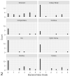
The histogram shows the bacterial index grade for inflammatory lesions split by tissue in 24 cases of feline ocular mycobacteriosis.
Figures 3–10.
Figures 3–10.
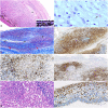
Mycobacteriosis, eye, cat. Inflammation scores: 1 = less than 1000 inflammatory cells; 2 = 1000–10,000 inflammatory cells; 3 = 10,001–50,000 inflammatory cells; 4 = 50,001–100,000 inflammatory cells; 5 = more than 100,000 inflammatory cells, calculated over the entire area of the lesion. Figure 3. Choroid, Mycobacterium bovis, case 7. Pyogranulomatous choroiditis, inflammation score 5. There is full-thickness infiltration of the choroid with inflammatory cells. Necrosis and edema are also present (asterisk). There is hypertrophy of the retinal pigment epithelium, indicating pathological retinal detachment (inset). Hematoxylin and eosin (HE). Figure 4. Choroid, Mycobacterium microti, case 2. Pyogranulomatous choroiditis, inflammation score 4, with evidence of acid-fast bacilli, some of which display the S-shaped morphology suggestive of M. microti (black arrow). Bacterial index grade 4. Ziehl-Neelsen (ZN). Figure 5. Choroid, M. bovis, case 1. Choroidal fibrosis is demonstrated by the presence of collagen fibers within the choroid (black arrows). Note the more densely arranged collagen fibers of the sclera (red arrows) compared with the collagen fibers within the choroid. Inflammation score 5. Masson’s trichrome. Figure 6. Choroid, Mycobacterium tuberculosis-complex (MTBC), case 13. Pyogranulomatous choroiditis, inflammation score 5, demonstrating abundant positive immunohistochemical labeling for ionized calcium-binding adaptor protein-1 (Iba1) on epithelioid macrophages, and lack of positivity on neutrophils and lymphocytes. Immunohistochemistry (IHC) for Iba1. Figure 7. Choroid, M. bovis, case 4. Inflammation score 5. Positive membranous labeling for T cells, forming the lymphocytic cuff in a granuloma. IHC for CD3. Figure 8. Choroid, M. bovis, case 17. Inflammation score 5. Large numbers of B cells infiltrating the choroid in a case of mixed pyogranulomatous and lymphoplasmacytic choroiditis. IHC for Pax5. Figure 9. Retina, MTBC, case 13. Macrophages and neutrophils are observed within the retina, with a lesser lymphoplasmacytic infiltrate. HE. Figure 10. Retina, M. bovis, case 18. Mixed pyogranulomatous and lymphoplasmacytic retinitis, inflammation score 3. Iba1-positive cells present within the inner layers of the retina, some of which have a more spindle-shaped morphology suggestive of resident microglia (black arrows). IHC for Iba1.
Figures 11–18.
Figures 11–18.
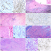
Mycobacteriosis, eye, cat. Inflammation scores: 1 = less than 1000 inflammatory cells; 2 = 1000–10,000 inflammatory cells; 3 = 10,001–50,000 inflammatory cells; 4 = 50,001–100,000 inflammatory cells; 5 = more than 100,000 inflammatory cells, calculated over the entire area of the lesion. Figure 11. Iris, Mycobacterium bovis, case 5. Inflammation score 2. (a) Lymphoplasmacytic iritis with scant T cells scattered throughout the iris stroma (black arrow). Immunohistochemistry (IHC) for CD3. (b) B cells are more abundant and outnumber T cells. IHC for Pax5. Figure 12. Ciliary body, M. bovis, case 9. Pyogranulomatous cyclitis, inflammation score 5, with extensive necrosis (asterisk) and loss of the epithelium of the pars plicata. There is a cyclitic membrane (black arrow), and Morgagnian globules in the lens (inset), indicating a cataract. Hematoxylin and eosin (HE). Figure 13. Sclera and episclera, M. bovis, case 4. Pyogranulomatous scleritis, inflammation score 5, with degeneration of collagen (asterisk) and a cluster of lymphocytes (black arrow). HE. Figure 14. Sclera, Mycobacterium lepraemurium, case 22. Pyogranulomatous scleritis, inflammation score 4, with “atypical” granulomas showing immunolabeled monocytes and granulocytes, mostly within the granuloma; macrophages and epithelioid macrophages are negative. IHC for calprotectin. Figure 15. Corneal limbus, M. lepraemurium, case 21. Inflammation score 3. (a) Pyogranulomatous perilimbal inflammation expanding the stroma, with the formation of non-necrotic pyogranulomas dominated by epithelioid macrophages. Normal features of the corneal limbus such as blood vessels (black arrow) and melanocytes are present. HE. (b) Positive staining for acid-fast bacilli indicative of mycobacteria within pyogranulomas. Bacterial index grade 5. Ziehl-Neelsen (ZN). Figure 16. Conjunctiva, Mycobacterium microti, case 3. Pyogranulomatous conjunctivitis of the third eyelid, inflammation score 5. Collagen fibers are present, subdividing the inflammatory infiltrate. The fibrous capsules are thin and incomplete. Masson’s trichrome. Figure 17. Optic nerve, M. bovis, case 8. Inflammation score 5. (a) Pyogranulomatous optic neuritis with necrosis (asterisk). HE. (b) Abundant acid-fast bacilli (pink) present within the optic nerve. The overall bacterial index grade was 5. ZN. Figure 18. Vitreous, M. bovis, case 17. Neutrophils are numerous within the vitreous. IHC for calprotectin.

Similar articles

References

    1. Agarwal A, Agrawal R, Gunasekaran DV, et al.. The Collaborative Ocular Tuberculosis Study (COTS)-1 Report 3: polymerase chain reaction in the diagnosis and management of tubercular uveitis: global trends. Ocul Immunol Inflamm. 2019;27:465–473. - PubMed
    1. Agostinelli C, Sabattini E, Gjørret JO, et al.. Characterization of a new monoclonal antibody against Pax5/BASP in 1525 paraffin-embedded human and animal tissue samples. Appl Immunohistochem Mol Morphol. 2010;18:561–572. - PubMed
    1. Aguilar JP, Green WR. Choroidal rupture. A histopathologic study of 47 cases. Retina. 1984;4:269–275. - PubMed
    1. Albert DM, Raven ML. Ocular tuberculosis. Microbiol Spectr. 2016;4. doi:10.1128/microbiolspec.TNMI7-0001-2016 - PMC - PubMed
    1. Anderson DH, Stern WH, Fisher SK, et al.. Retinal detachment in the cat: the pigment epithelial-photoreceptor interface. Invest Ophthalmol Vis Sci. 1983;24:906–926. - PubMed

Publication types

Substances