. 2022 May;13(5):12532-12547.
doi: 10.1080/21655979.2022.2076497.

Integrin-linked kinase affects the sensitivity of esophageal squamous cell carcinoma cells to chemotherapy with cisplatin via the Wnt/beta-catenin signaling pathway

Affiliations

Integrin-linked kinase affects the sensitivity of esophageal squamous cell carcinoma cells to chemotherapy with cisplatin via the Wnt/beta-catenin signaling pathway

Ting Xu et al. Bioengineered. 2022 May.

Abstract

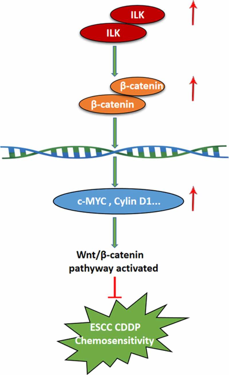
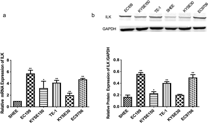
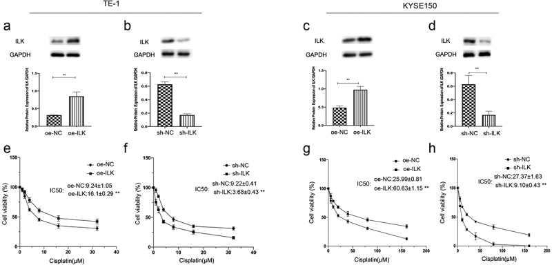
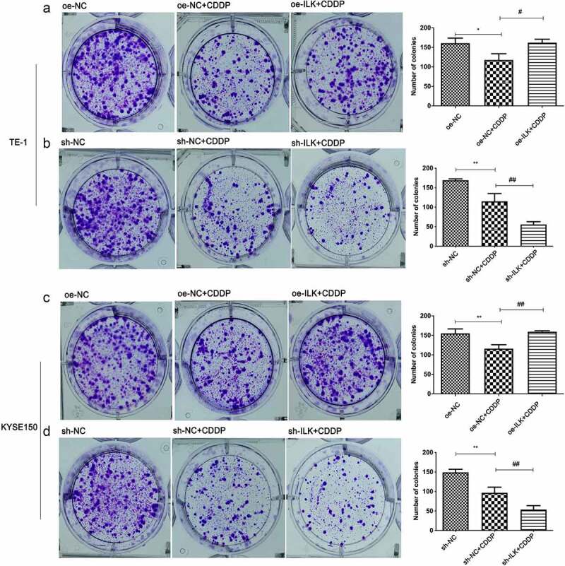
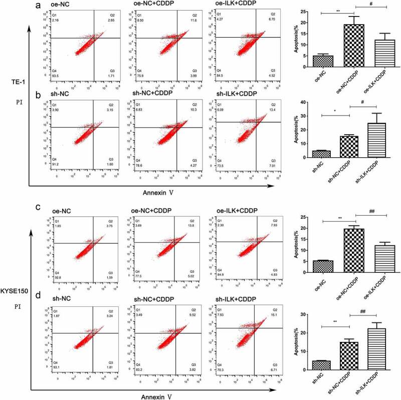
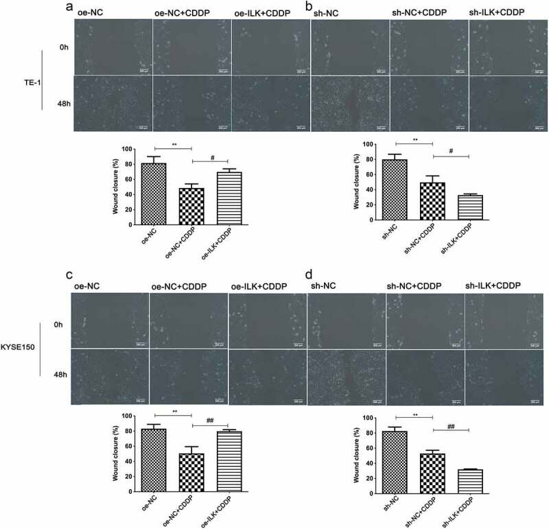
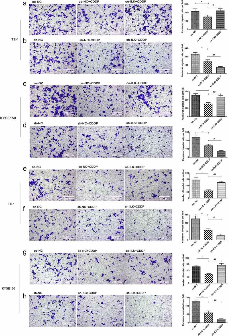
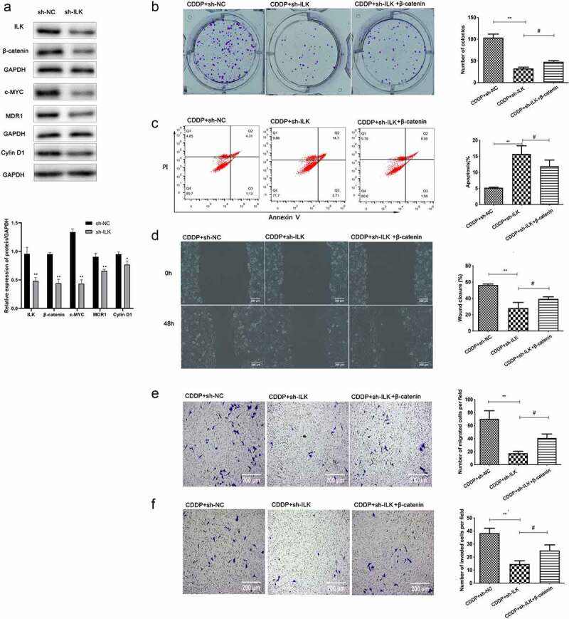
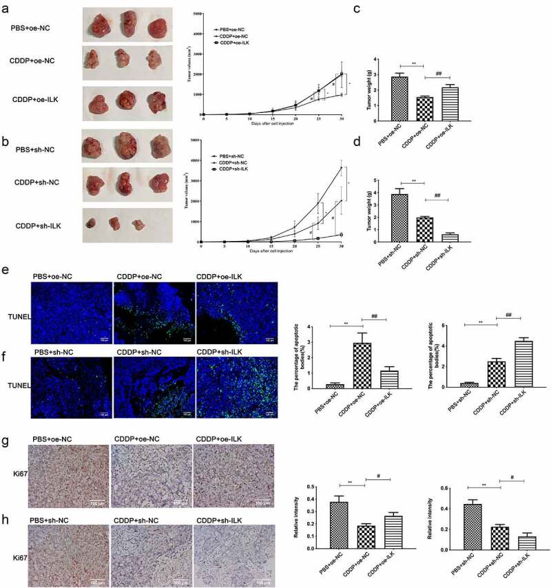
Recent studies have shown that the expression of integrin-linked kinase (ILK) was related to the occurrence, development, and malignant progression of esophageal squamous cell carcinoma (ESCC). However, research on the relationship between ILK and the chemosensitivity of ESCC has to date not been reported. The present study found that ILK was highly expressed in ESCC cell lines, and the overexpression of ILK in ESCC cells reduced the incidence of cell apoptosis and alleviated the cytotoxicity on cells induced by cisplatin (CDDP). Inversely, ILK knockdown increased CDDP-induced apoptosis and had an inhibitive effect on the malignant phenotype of ESCC, including cell proliferation, invasion, and migration. In addition, ILK knockdown in ESCC cells inhibited the expression of beta (β)-catenin and activated the wingless/integrated (Wnt) signaling pathway. Furthermore, cellular MYC (c-MYC) and Cylin D1 were the target genes of the Wnt signaling pathway. Rescue experiments showed that the overexpression of β-catenin reversed a tumor's inhibition and apoptosis abilities induced by ILK knockdown. In conclusion, ILK potentially reduced the CDDP sensitivity of ESCC cells by influencing the activity of the Wnt/β-catenin signaling pathway.


Conflict of interest statement

No potential conflict of interest was reported by the author(s).

Figures

None
Graphical abstract
Figure 1.
Figure 1.
The expression level of ILK in ESCC cells. (a) ILK detected by RT-PCR in five ESCC cell lines and one esophageal epithelial cell line (SHEE). (b)WB analysis of ILK expression in ESCC cell lines and SHEE.*p < 0.05,**p < 0.01.
Figure 2.
Figure 2.
ILK affects ESCC sensitivity to DDP. (a-d) Relative expression of ILK was assayed in KYSE150 and TE-1 cells infected with oe-ILK(overexpression of ILK) or sh-ILK (konckdown of ILK) by WB. (e-f) The IC50 of TE-1 to CDDP after transfection with oe-ILK and sh-ILK. (g-h) The IC50 of KYSE150 to CDDP after transfection with oe-ILK and sh-ILK. *p < 0.05, **p < 0.01.
Figure 3.
Figure 3.
Effects of ILK on the proliferation of ESCC cells toward CDDP. (a-b) Proliferative capacity of TE-1 exposed to DDP after transfection with oe-ILK and sh-ILK was analyzed by colony formation assay. (c-d) Proliferative capacity of KYSE150 exposed to DDP after transfection with oe-ILK and sh-ILK was analyzed by colony formation assay.*#p < 0.05 and **##p < 0.01.
Figure 4.
Figure 4.
Effection of ILK on apoptosis of ESCC cells toward to CDDP. (a-b) Cell apoptosis of TE-1 induced by CDDP after transfection with oe-ILK and sh-ILK was analyzed with flow cytometry. (c-d) Cell apoptosis of KYSE150 induced by CDDP after transfection with oe-ILK and sh-ILK was analyzed by flow cytometry. #p < 0.05 and **##p < 0.01.
Figure 5.
Figure 5.
Effects of ILK on ESCC cell migration to CDDP. (a-b) Effect of ILK on cell migration toward CDDP was assessed by wound healing assays in TE-1. (c-d) Effect of ILK on cell migration toward CDDP was assessed by wound healing assays in KYSE150. #p < 0.05 and **##p < 0.01.
Figure 6.
Figure 6.
Effects of ILK on ESCC cell migration and invasion to CDDP. (a-b) Effect of ILK on cell migration toward CDDP was assessed by Transwell assays without Matrigel in TE-1. (c-d) Effect of ILK on cell migration toward CDDP was assessed by Transwell assays without Matrigel in KYSE150. (e-f) Effect of ILK on cell invasion toward CDDP was assessed by Transwell assays with Matrigel in TE-1. (g-h) Effect of ILK on cell invasion toward CDDP was assessed by Transwell assays with Matrigel in KYSE150. *#p < 0.05 and **p < 0.01.
Figure 7.
Figure 7.
Knockdown of ILK promotes the sensitivity of ESCC cells to CDDP through disturbing the expression of β-catenin. (a) The expression level of ILK, β-catenin, c-MYC, MRD1 Cylin D1, level was detected by WB in TE-1 cells after transfected with sh-ILK and sh-NC. (b) Effects of β-catenin overexpression on cell colony formation to CDDP after knockdown of ILK. (c) Effects of β-catenin overexpression on cell apoptosis to CDDP after knockdown of ILK. (d-e) Effects of β-catenin overexpression on cell migration to CDDP after knockdown of ILK by wound healing assays and Transwell assays (without Matrigel). (f) Effects of β-catenin overexpression on cell invasion to CDDP after knockdown of ILK by transwell assays (with Matrigel).* #p < 0.05 and **p < 0.01.
Figure 8.
Figure 8.
Effects of ILK expression on the xenograft tumor formation by ESCC cancer cells in vivo toward CDDP treatment. (a-b) The tumor volume of each group. (c-d)The tumor weight from the xenografts all mice were killed at the end of the experiment. (e) Cell apoptosis in each group was assessed by TUNEL (f) The expression of cell proliferation related protein Ki67 in each group was detected by Immunohistochemistry. *#p < 0.05 and **##p < 0.01.

Similar articles

Cited by

References

    1. Sung H, Ferlay J, Siegel RL, et al. Global cancer statistics 2020: GLOBOCAN estimates of incidence and mortality worldwide for 36 cancers in 185 countries [J]. CA Cancer J Clin. 2021:1–41. - PubMed
    1. Ohashi S, Miyamoto S, Kikuchi O, et al. Recent Advances From Basic and Clinical Studies of Esophageal Squamous Cell Carcinoma. Gastroenterology. 2015;149:1700–1715. - PubMed
    1. Pennathur A, Gibson MK, Jobe BA, et al. Oesophageal carcinoma. Lancet. 2013;381:400–412. - PubMed
    1. Hanahan D, Weinberg RA.. Hallmarks of cancer: the next generation. Cell. 2011;144:646–674. - PubMed
    1. Chan D, Zhou Y, Chui CH, et al. Expression of insulin-like growth factor binding protein-5 (IGFBP5) reverses cisplatin-resistance in esophageal carcinoma. Cells. 2018;7(10):143. - PMC - PubMed

MeSH terms