. 2022 May 19;18(5):e1010498.
doi: 10.1371/journal.ppat.1010498. eCollection 2022 May.

Clofoctol inhibits SARS-CoV-2 replication and reduces lung pathology in mice

Affiliations

Clofoctol inhibits SARS-CoV-2 replication and reduces lung pathology in mice

Sandrine Belouzard et al. PLoS Pathog. .

Abstract

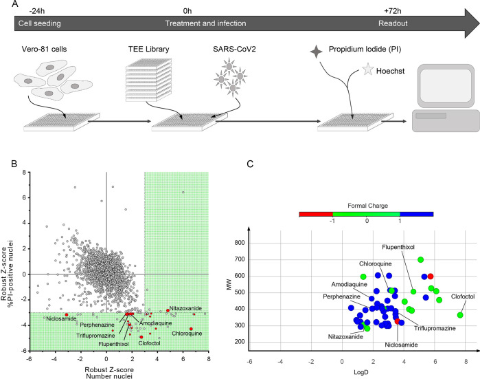
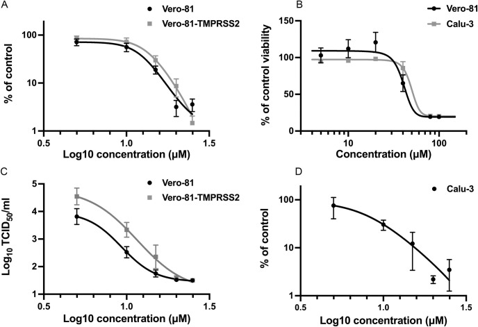
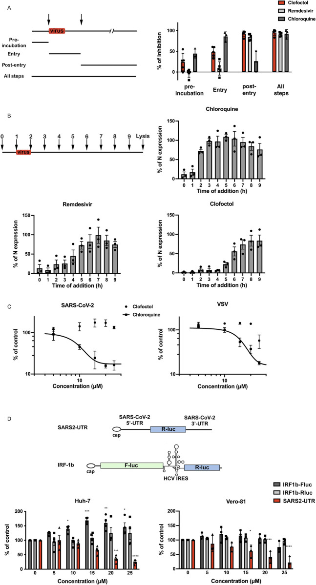
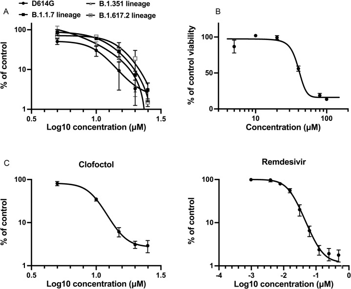
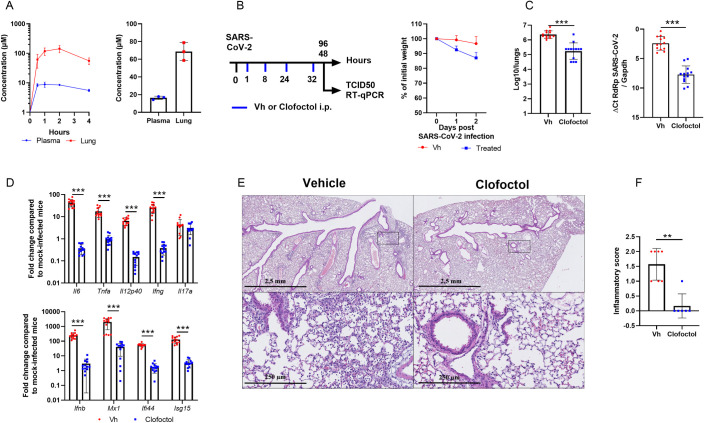
Drug repurposing has the advantage of shortening regulatory preclinical development steps. Here, we screened a library of drug compounds, already registered in one or several geographical areas, to identify those exhibiting antiviral activity against SARS-CoV-2 with relevant potency. Of the 1,942 compounds tested, 21 exhibited a substantial antiviral activity in Vero-81 cells. Among them, clofoctol, an antibacterial drug used for the treatment of bacterial respiratory tract infections, was further investigated due to its favorable safety profile and pharmacokinetic properties. Notably, the peak concentration of clofoctol that can be achieved in human lungs is more than 20 times higher than its IC50 measured against SARS-CoV-2 in human pulmonary cells. This compound inhibits SARS-CoV-2 at a post-entry step. Lastly, therapeutic treatment of human ACE2 receptor transgenic mice decreased viral load, reduced inflammatory gene expression and lowered pulmonary pathology. Altogether, these data strongly support clofoctol as a therapeutic candidate for the treatment of COVID-19 patients.


Conflict of interest statement

I have read the journal’s policy and the authors of this manuscript have the following competing interests:European Patent Application Serial No. EP20305633.8, entitled “Compound and method for the treatment of coronaviruses” related to this work was filed on 10 June 2020. Authors TB, LB, CM, SB, PB, ND, BD, JeD, EH, AM, YR and TV of this manuscript are inventors of the patent.

Figures

Fig 1
Fig 1. HCS screen of Apteeus TEElibrary for the identification of anti-SARS-CoV-2 compounds.
A, Workflow overview of the screening process. B, Dot-plot representations of all compounds tested based on their robust-Z-score for both the numbers of nuclei and the percentages of PI-positive nuclei. Dotted lines are indicative of the thresholds chosen for hit selectivity (within the green area). C, Dot-plot representation according to the molecular weight and the LogD of the compounds of interest. Dots are color-coded based on the ionization state at physiological pH.
Fig 2
Fig 2. In vitro validation of the antiviral activity of clofoctol.
A, Clofoctol inhibits the genomic replication of SARS-CoV-2. Vero-81 and Vero-81-TMPRSS2 cells were infected for 6h at an MOI of 0.25 in the presence of increasing concentrations of clofoctol. Then, total RNA was extracted and viral RNA was quantified by RT-qPCR and normalized by the amount of total RNA. Results are presented as the percentage of the viral load of the control and represent the average of seven independent experiments performed in duplicates. Error bars represent the standard error of the mean (SEM). B, Clofoctol is not cytotoxic in cell culture at concentrations below 40 μM. Vero-81 cells and Calu-3 cells were cultured in the presence of given concentrations of clofoctol. Cell viability was monitored using the MTS-based viability assay after 24 hours of incubation. C, Clofoctol inhibits the production of progeny virions. Vero-81 and Vero-81-TMPRSS2 cells were infected with SARS-CoV-2 at a MOI of 0.25. After 1h, the inoculum was removed and the cells were washed with PBS prior treatment with clofoctol. Cells were then further incubated for 16h. Thereafter, supernatants were collected and the amounts of secreted infectious virus were quantified. The limit of detection was 1.5TCID50/mL. These data represent the average of three independent experiments (N = 3). Experiments were performed in duplicate for each condition. D, Clofoctol inhibits SARS-CoV-2 replication in Calu-3 cells. Calu-3 cells were infected at a MOI of 0.25 in the presence of increasing concentrations of clofoctol for 24h. Then, total cellular RNA was extracted and viral RNA was quantified by RT-qPCR. Results are presented as the percentage of the viral load of the control and represent the average of three independent experiments performed in duplicates. Error bars represent the standard error of the mean (SEM).
Fig 3
Fig 3. Clofoctol targets the translation step of SARS-CoV-2 life cycle.
A, Clofoctol is mainly efficient at a post-entry step. Vero-81 cells were infected with SARS-CoV-2 at an MOI of 0.25. Clofoctol, remdesivir or CQ were present at a concentration of 15 μM either before infection, during virus entry, post-inoculation or throughout the steps as indicated on the schematic depiction of the experiment. Bars indicate when the drugs are present during the experiment for each condition. At 16h post-infection, cells were fixed with 4% paraformaldehyde and processed to detect the proportion of infected cells. Therefore, they were immunostained to allow for the detection of the viral double-stranded RNA and nuclei were detected by Hoechst staining to count the total number of cells. Results are presented as the percentage of infection inhibition and represent the average of five independent experiments. B, Time-of-addition experiment. Vero-81 cells were infected at an MOI of 0.5 for 1h. 15 μM of clofoctol, remdesivir or CQ were added every hour starting 1h before inoculation. Cells were lysed 8h after the end of the inoculation in Laemmli loading buffer and the amount of N protein was detected in immunoblot. Results are presented as the percentage of N protein expression relative to that in non-treated cells (CTL) and represent the average of three independent experiments. Error bars represent the standard error of the mean (SEM). C, Clofoctol does not inhibit SARS-CoV-2 entry. Huh-7 cells expressing ACE2 receptor were infected with SARS2pp or pseudoparticles containing the envelope glycoprotein of the vesicular stomatitis virus (VSV) used as a control (VSVpp) for 3 hours in the presence of increasing concentrations of clofoctol or CQ. At 48 hours post-infection, cells were lysed to quantify luciferase activity. The results are expressed in % of the controls of three independent experiments. The experiments were performed in triplicate (n = 3) in each condition. D, Clofoctol inhibits viral RNA translation. Schematic representation of the reporter construct expressing the Renilla luciferase placed between the 5’-UTR and the 3’-UTR of the SARS-CoV-2 genomic RNA and the control bicistronic construct containing the firefly luciferase sequence under the control of a cap structure, followed by the Renilla luciferase under the control of hepatitis C virus (HCV) IRES. Huh-7 or Vero-81 cells were electroporated with in vitro transcribed RNA. Cells were lysed after 8h and luciferase activities were recorded. The results are expressed in % of the controls of three independent experiments. The experiments were performed in quadruplicate (n = 4) in each condition. Two-way ANOVA followed by the Dunnett’s multiple comparisons test was performed for statistical analysis (*p < 0.05; **p < 0.01; ***p < 0.001).
Fig 4
Fig 4. Clofoctol inhibits other SARS-CoV-2 variants as well as HCoV-229E.
A, Vero-81 cells were infected either with SARS-CoV-2 of lineage B1 containing the D614G mutation (SARS-CoV-2/human/FRA/Lille_Vero-TMPRSS2/2020) or with SARS-CoV-2 of lineage B1.1.7 (GISAID accession number EPI_ISL_1653931) or lineage B.1.351 (GISAID accession number EPI_ISL_1653932) or lineage B.1.617.2 (GISAID accession number EPI_ISL_2143633). Viral genomes were quantified by RT-qPCR and normalized by the amount of total RNA. Results are presented as the percentage of the viral load of the control and represent the average of three independent experiments performed in duplicates. Error bars represent the standard error of the mean (SEM). B, Clofoctol is not cytotoxic in cell culture at concentrations below 40 μM. Huh-7 cells were cultured in the presence of given concentrations of clofoctol. Cell viability was monitored using the MTS-based viability assay after 24 hours of incubation. C, Huh-7 cells were infected with HCoV-229E-Rluc in presence of different concentrations of clofoctol or remdesivir. At 7h post-infection, cells were lysed and luciferase activities were quantified. Results are presented as the percentages of the control and represent an average of three independent experiments performed in triplicates. Errors bars represent the standard error of the mean (SEM).
Fig 5
Fig 5. Pharmacokinetics and antiviral properties of clofoctol in a mouse model of COVID-19.
A, Pharmacokinetics characterization of clofoctol in mice. Left panel, 8–10 week-old female C57BL/6J mice were treated i.p. with a single dose of clofoctol (62.5mg/kg) and were sacrificed at different time points thereafter. Right panel, Clofoctol was inoculated twice daily during two days and mice were sacrificed 1h after the last injection. Clofoctol concentrations in lungs (n = 3/time point, 3 samples/lung) and plasma (n = 3/time point, 2 technical replicates) are depicted. B-D, Effects of clofoctol treatment on SARS-CoV-2 infection in K18-hACE2 transgenic C57BL/6J mice. B, Left panel, Scheme of the experimental design in which the effect of clofoctol was assessed in mice. Mice were treated i.p. with clofoctol (62.5mg/kg) or vehicle 1h and 8h after i.n. inoculation of SARS-CoV-2 (5x10² TCID50 per mouse) and treated again twice at day 1 post-infection. Animals were sacrificed at day 2 and day 4 post-infection. Right panel, Body weight curves are shown. C,The viral load was determined by titration on Vero-E6 cells (middle panel) and by RT-qPCR (right panel) (day 2 post-infection). D, mRNA copy numbers of genes were quantified by RT-qPCR. Data are expressed as fold change over average gene expression in mock-treated (uninfected) animals (day 2 post-infection). E, Lung sections were analyzed at day 4 post-infection. Shown are representative lungs (hematoxylin and eosin staining). Lower panels, enlarged views of the area circled in black in upper panels. F, Blinded sections were scored for levels of pathological severity. The inflammatory score is depicted. B-C, Results are expressed as the mean ± SD (n = 13 for panels B and D and n = 6–7 for panel E and F). Significant differences were determined using the Mann-Whitney U test (**p < 0.01; ***p < 0.001).

Similar articles

Cited by

  • A community effort in SARS-CoV-2 drug discovery.
    Schimunek J, Seidl P, Elez K, Hempel T, Le T, Noé F, Olsson S, Raich L, Winter R, Gokcan H, Gusev F, Gutkin EM, Isayev O, Kurnikova MG, Narangoda CH, Zubatyuk R, Bosko IP, Furs KV, Karpenko AD, Kornoushenko YV, Shuldau M, Yushkevich A, Benabderrahmane MB, Bousquet-Melou P, Bureau R, Charton B, Cirou BC, Gil G, Allen WJ, Sirimulla S, Watowich S, Antonopoulos N, Epitropakis N, Krasoulis A, Itsikalis V, Theodorakis S, Kozlovskii I, Maliutin A, Medvedev A, Popov P, Zaretckii M, Eghbal-Zadeh H, Halmich C, Hochreiter S, Mayr A, Ruch P, Widrich M, Berenger F, Kumar A, Yamanishi Y, Zhang KYJ, Bengio E, Bengio Y, Jain MJ, Korablyov M, Liu CH, Marcou G, Glaab E, Barnsley K, Iyengar SM, Ondrechen MJ, Haupt VJ, Kaiser F, Schroeder M, Pugliese L, Albani S, Athanasiou C, Beccari A, Carloni P, D'Arrigo G, Gianquinto E, Goßen J, Hanke A, Joseph BP, Kokh DB, Kovachka S, Manelfi C, Mukherjee G, Muñiz-Chicharro A, Musiani F, Nunes-Alves A, Paiardi G, Rossetti G, Sadiq SK, Spyrakis F, Talarico C, Tsengenes A, Wade RC, Copeland C, Gaiser J, Olson DR, Roy A, Venkatraman V, Wheeler TJ, Arthanari H, Blaschitz K, Cespugli M, Durmaz V, Fackeldey K, Fischer PD, Gorgulla C, Gruber C, Gruber K, Hetmann M, Kin… See abstract for full author list ➔ Schimunek J, et al. Mol Inform. 2024 Jan;43(1):e202300262. doi: 10.1002/minf.202300262. Epub 2023 Nov 14. Mol Inform. 2024. PMID: 37833243 Free PMC article.
  • Discovery of Anti-Coronavirus Cinnamoyl Triterpenoids Isolated from Hippophae rhamnoides during a Screening of Halophytes from the North Sea and Channel Coasts in Northern France.
    Al Ibrahim M, Akissi ZLE, Desmarets L, Lefèvre G, Samaillie J, Raczkiewicz I, Sahpaz S, Dubuisson J, Belouzard S, Rivière C, Séron K. Al Ibrahim M, et al. Int J Mol Sci. 2023 Nov 22;24(23):16617. doi: 10.3390/ijms242316617. Int J Mol Sci. 2023. PMID: 38068938 Free PMC article.
  • Hyperforin, the major metabolite of St. John's wort, exhibits pan-coronavirus antiviral activity.
    Raczkiewicz I, Rivière C, Bouquet P, Desmarets L, Tarricone A, Camuzet C, François N, Lefèvre G, Silva Angulo F, Robil C, Trottein F, Sahpaz S, Dubuisson J, Belouzard S, Goffard A, Séron K. Raczkiewicz I, et al. Front Microbiol. 2024 Aug 8;15:1443183. doi: 10.3389/fmicb.2024.1443183. eCollection 2024. Front Microbiol. 2024. PMID: 39176276 Free PMC article.
  • A reporter cell line for the automated quantification of SARS-CoV-2 infection in living cells.
    Desmarets L, Callens N, Hoffmann E, Danneels A, Lavie M, Couturier C, Dubuisson J, Belouzard S, Rouillé Y. Desmarets L, et al. Front Microbiol. 2022 Sep 29;13:1031204. doi: 10.3389/fmicb.2022.1031204. eCollection 2022. Front Microbiol. 2022. PMID: 36246297 Free PMC article.
  • Lichen or Associated Micro-Organism Compounds Are Active against Human Coronaviruses.
    Desmarets L, Millot M, Chollet-Krugler M, Boustie J, Camuzet C, François N, Rouillé Y, Belouzard S, Tomasi S, Mambu L, Séron K. Desmarets L, et al. Viruses. 2023 Aug 31;15(9):1859. doi: 10.3390/v15091859. Viruses. 2023. PMID: 37766264 Free PMC article.

References

    1. Arthi V, Parman J. Disease, downturns, and wellbeing: Economic history and the long-run impacts of COVID-19. Explor Econ Hist. 2021;79: 101381. doi: 10.1016/j.eeh.2020.101381 - DOI - PMC - PubMed
    1. WHO Solidarity Trial Consortium, Pan H, Peto R, Henao-Restrepo A-M, Preziosi M-P, Sathiyamoorthy V, et al.. Repurposed Antiviral Drugs for Covid-19—Interim WHO Solidarity Trial Results. N Engl J Med. 2021;384: 497–511. doi: 10.1056/NEJMoa2023184 - DOI - PMC - PubMed
    1. Jeon S, Ko M, Lee J, Choi I, Byun SY, Park S, et al.. Identification of Antiviral Drug Candidates against SARS-CoV-2 from FDA-Approved Drugs. Antimicrob Agents Chemother. 2020;64. doi: 10.1128/AAC.00819-20 - DOI - PMC - PubMed
    1. Riva L, Yuan S, Yin X, Martin-Sancho L, Matsunaga N, Pache L, et al.. Discovery of SARS-CoV-2 antiviral drugs through large-scale compound repurposing. Nature. 2020;586: 113–119. doi: 10.1038/s41586-020-2577-1 - DOI - PMC - PubMed
    1. Yuan S, Yin X, Meng X, Chan JF-W, Ye Z-W, Riva L, et al.. Clofazimine broadly inhibits coronaviruses including SARS-CoV-2. Nature. 2021. doi: 10.1038/s41586-021-03431-4 - DOI - PubMed

Publication types