. 2022 May 19;17(5):e0268626.
doi: 10.1371/journal.pone.0268626. eCollection 2022.

Transcriptome analysis reveals high tumor heterogeneity with respect to re-activation of stemness and proliferation programs

Affiliations

Transcriptome analysis reveals high tumor heterogeneity with respect to re-activation of stemness and proliferation programs

Artem Baranovsky et al. PLoS One. .

Abstract

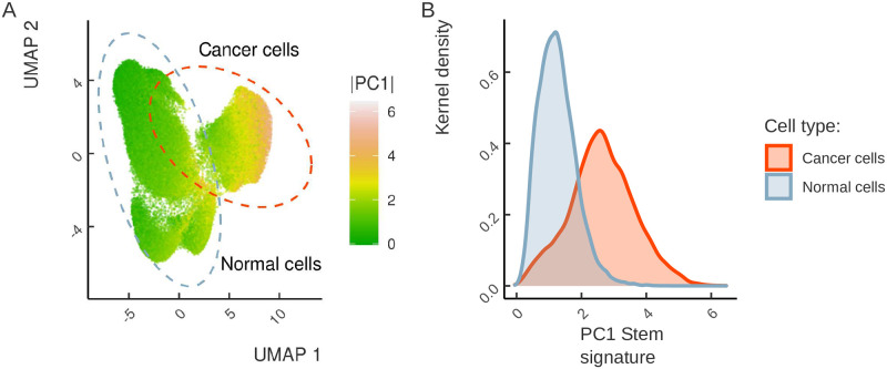
Significant alterations in signaling pathways and transcriptional regulatory programs together represent major hallmarks of many cancers. These, among all, include the reactivation of stemness, which is registered by the expression of pathways that are active in the embryonic stem cells (ESCs). Here, we assembled gene sets that reflect the stemness and proliferation signatures and used them to analyze a large panel of RNA-seq data from The Cancer Genome Atlas (TCGA) Consortium in order to specifically assess the expression of stemness-related and proliferation-related genes across a collection of different tumor types. We introduced a metric that captures the collective similarity of the expression profile of a tumor to that of ESCs, which showed that stemness and proliferation signatures vary greatly between different tumor types. We also observed a high degree of intertumoral heterogeneity in the expression of stemness- and proliferation-related genes, which was associated with increased hazard ratios in a fraction of tumors and mirrored by high intratumoral heterogeneity and a remarkable stemness capacity in metastatic lesions across cancer cells in single cell RNA-seq datasets. Taken together, these results indicate that the expression of stemness signatures is highly heterogeneous and cannot be used as a universal determinant of cancer. This calls into question the universal validity of diagnostic tests that are based on stem cell markers.


Conflict of interest statement

The authors have declared that no competing interests exist.

Figures

Fig 1
Fig 1. The stemness signature gene set intersects with previously published ESC signature gene sets.
The number of genes in the intersection between sets on the X and Y axes is shown in the cells of the matrix. The color scale encodes the size of the intersection relative to the size of the dataset on the Y axis (above the diagonal) and relative to the size of the dataset on the X axis (below the diagonal).
Fig 2
Fig 2. Principal Component Analysis (PCA) of the expression of stemness signatures (A), proliferation signatures (B), and genes representing EMT-MET (C) in differentiating iPSCs, ESCs, ASCs, tumors and normal tissues shown as individual samples (top) and density (bottom).
Tumors span continuously between normal tissues and iPSCs in the expression subspaces of stemness, proliferation, but not EMT-MET genes. Tumor samples form two clusters: one closer to ESCs and iPSCs, another closer to the normal tissues and ASCs.
Fig 3
Fig 3. Heterogeneity of stemness and proliferation signatures across tumor types.
The first principal component (PC1) encodes the signature of stemness (A), proliferation (B), and EMT-MET signature genes (C). The samples are plotted separately for each tumor in the TCGA project, normal tissues, and iPSCs. Tumor samples are colored according to their position relative to the PC1 median (black vertical line). Tumor samples located to the right (left) of the median are assigned to the Normal (Stem) cluster, respectively. (D) The standard TCGA notation for cancer types.
Fig 4
Fig 4. The relationship between stemness and proliferation intensities.
(A) Stemness intensity vs. proliferation intensity in 19 solid tumors with different average project-wise tumor stage and median project-wise purity. (B) A heatmap of stemness and proliferation intensities across 19 solid tumors from TCGA. Two groups of tumor samples are identified: top-right quadrant (stemness Intensity>0.5 and proliferation Intensity>0.5) and bottom-left quadrant (stemness Intensity<0.5 and proliferation Intensity<0.5). The notation for cancer types as in Fig 3. Tumors from stem cell rich organs are marked with asterisks.
Fig 5
Fig 5. Intertumor heterogeneity of stemness and proliferation signatures.
(A) Stem and Normal clusters specific to each tumor type are defined by the location of a sample relative to the median PC1 value across all samples of the given tumor (plotted as horizontal black lines). Samples above (below) the median are assigned to the Normal (Stem) cluster, respectively. (B) Hazard ratios for the comparison of patient survival in Stem and Normal clusters in two sample groups (top-right and bottom-left in Fig 4) with respect to stemness and proliferation intensity. Error bars denote 95% confidence intervals. Tumor types are annotated at the bottom according to the average tumor stage and the median tumor purity. Codes for statistical significance reported are as follows: NS − p-value >0.05, * − 0.01< p-value <0.05, ** − 0.001< p-value <0.01, *** − p-value <0.001. The notation for cancer types as in Fig 3.
Fig 6
Fig 6. Intertumor heterogeneity of stemness and proliferation is predictive of poor survival.
Kaplan-Meier survival curves grouped by clustering according to stemness (proliferation) signature are plotted for skin melanomas (A), lung adenocarcinoma (B) and renal clear cell carcinoma (C). P-values from the logrank test are reported. The notation for cancer types as in Fig 3.
Fig 7
Fig 7. Cancer cells show substantial intratumor heterogeneity of stemness.
(A) UMAP dimensionality reduction of the colorectal cancer single-cell RNA-seq dataset. The annotation of normal and cancer cell clusters is from [38]. The cells are colored by the degree of stemness signature reactivation (the absolute value of PC1 projection). (B) The distribution of stemness signature (PC1) (X axis) in normal and cancer cells.
Fig 8
Fig 8. Cancer cells show heterogeneity in stemness, proliferation and EMT scores.
(A) Stemness, proliferation and EMT scores in single cells by cell type. Boxplots colors correspond to normal (blue), colorectal cancer (orange), and stem (red) cells. (B) Two dimensional embedding derived from UMAP colored by the stemness score from panel A. (C) Stemness scores computed for single cells from primary lung adenocarcinomas (orange), metastatic lesions (red), and surrounding normal tissues (blue). (D) Stemness scores of colorectal cancer cells from different patients.

References

    1. Roth GA, Abate D, Abate KHea. Global, regional, and national age-sex-specific mortality for 282 causes of death in 195 countries and territories, 1980-2017: a systematic analysis for the Global Burden of Disease Study 2017. Lancet. 2018;392(10159):1736–1788. doi: 10.1016/S0140-6736(18)32203-7 - DOI - PMC - PubMed
    1. Miller KD, Nogueira L, Mariotto AB, Rowland JH, Yabroff KR, Alfano CM, et al.. Cancer treatment and survivorship statistics, 2019. CA Cancer J Clin. 2019;69(5):363–385. doi: 10.3322/caac.21565 - DOI - PubMed
    1. Arruebo M, Vilaboa N, Sáez-Gutierrez B, Lambea J, Tres A, Valladares M, et al.. Assessment of the evolution of cancer treatment therapies. Cancers (Basel). 2011;3(3):3279–3330. doi: 10.3390/cancers3033279 - DOI - PMC - PubMed
    1. Baxevanis CN, Perez SA, Papamichail M. Combinatorial treatments including vaccines, chemotherapy and monoclonal antibodies for cancer therapy. Cancer Immunol Immunother. 2009;58(3):317–324. doi: 10.1007/s00262-008-0576-4 - DOI - PMC - PubMed
    1. Zhou BB, Zhang H, Damelin M, Geles KG, Grindley JC, Dirks PB. Tumour-initiating cells: challenges and opportunities for anticancer drug discovery. Nat Rev Drug Discov. 2009;8(10):806–823. doi: 10.1038/nrd2137 - DOI - PubMed

Publication types