

Attenuation of near-ultraviolet, visible and near-infrared light in sound and carious human enamel and dentin
- PMID: 35588022
- PMCID: PMC9474553
- DOI: 10.1007/s00784-022-04541-7
Attenuation of near-ultraviolet, visible and near-infrared light in sound and carious human enamel and dentin
Abstract
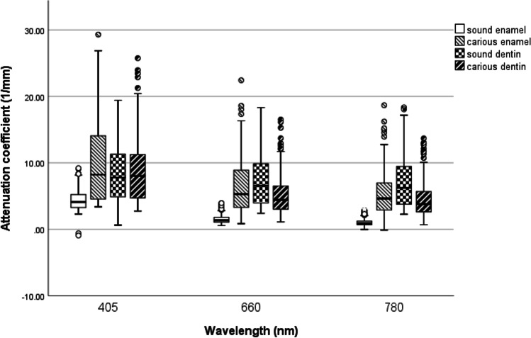
Objectives: This in vitro study aimed to investigate the optical attenuation of light at 405, 660 and 780 nm sent through sound and carious human enamel and dentin, including respective individual caries zones, as well as microscopically sound-appearing tissue close to a carious lesion.
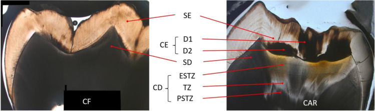
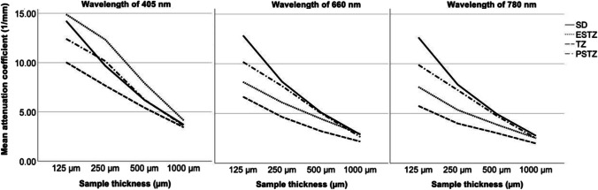
Materials and methods: Collimated light transmission through sections of 1000-125-µm thickness was measured and used to calculate the attenuation coefficient (AC). The data were statistically analysed with a MANOVA and Tukey's HSD. Precise definition of measurement points enabled separate analysis within the microstructure of lesions: the outer and inner halves of enamel (D1, D2), the translucent zone (TZ) within dentin lesions and its adjacent layers, the enamel side of the translucent zone (ESTZ) and the pulpal side of the translucent zone (PSTZ).
Results: The TZ could be distinguished from its adjacent layers and from caries-free dentin at 125 µm. Sound-appearing dentin close to caries lesions significantly differed from caries-free dentin at 125 µm. While sound and carious enamel exhibited a significant difference (p < 0.05), this result was not found for D1 and D2 enamel lesions (p > 0.05). At 405 nm, no difference was found between sound and carious dentin (p > 0.05).
Conclusions: Light optical means enable the distinction between sound and carious tissue and to identify the microstructure of dentin caries partially as well as the presence of tertiary dentin formation. Information on sample thickness is indispensable when interpreting the AC.
Clinical relevance: Non-ionising light sources may be suitable to detect lesion progression and tertiary dentin.
Keywords: Caries detection; Caries zones; Dentin caries; Enamel caries; Tertiary dentin; Transillumination.
© 2022. The Author(s).
Conflict of interest statement
The authors declare no competing interests.
Figures
References
-
- Kocak N, Cengiz-Yanardag E. Clinical performance of clinical-visual examination, digital bitewing radiography, laser fluorescence, and near-infrared light transillumination for detection of non-cavitated proximal enamel and dentin caries. Lasers Med Sci. 2020;35(7):1621–1628. doi: 10.1007/s10103-020-03021-2. - DOI - PubMed
MeSH terms
LinkOut - more resources
Full Text Sources
Medical
