. 2022 May 19;18(5):e1010171.
doi: 10.1371/journal.pgen.1010171. eCollection 2022 May.

MDM2 E3 ligase activity is essential for p53 regulation and cell cycle integrity

Affiliations

MDM2 E3 ligase activity is essential for p53 regulation and cell cycle integrity

Meenalakshmi Chinnam et al. PLoS Genet. .

Erratum in

Abstract

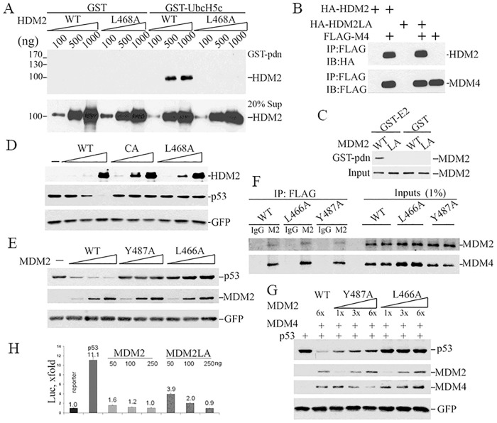
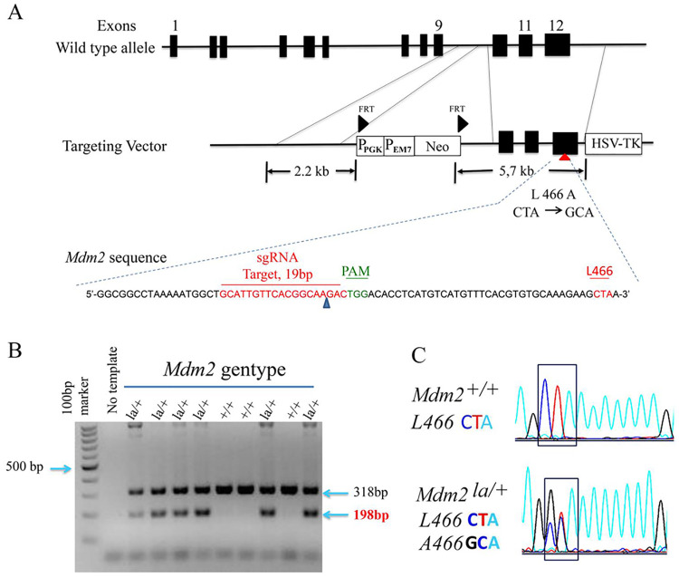
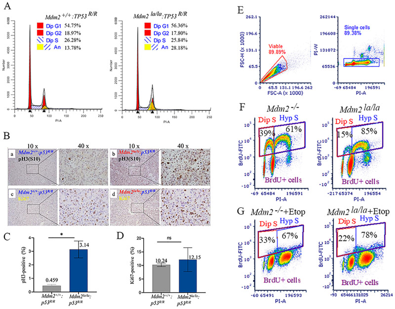
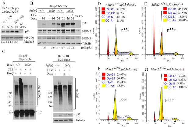
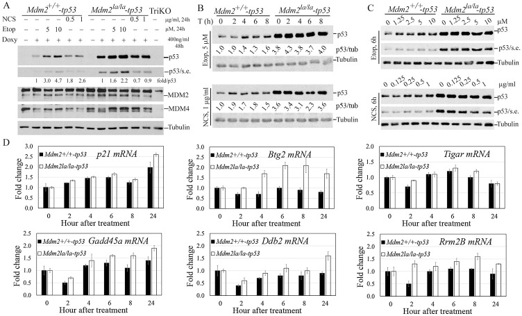
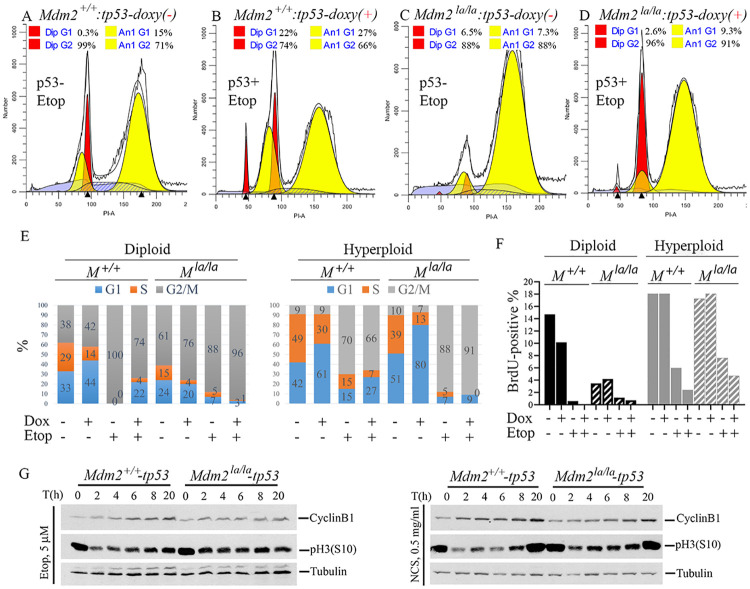
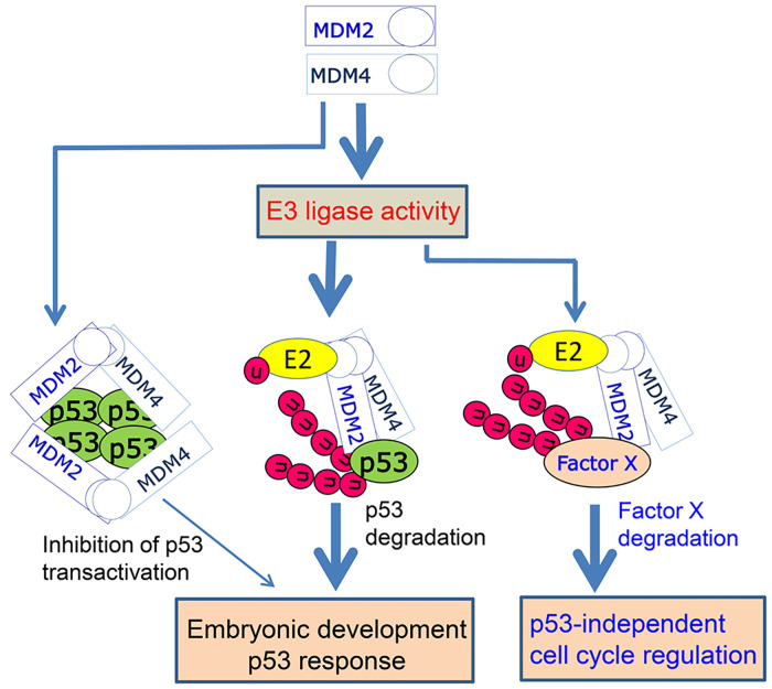
MDM2 and MDM4 are key regulators of p53 and function as oncogenes when aberrantly expressed. MDM2 and MDM4 partner to suppress p53 transcriptional transactivation and polyubiquitinate p53 for degradation. The importance of MDM2 E3-ligase-mediated p53 regulation remains controversial. To resolve this, we generated mice with an Mdm2 L466A mutation that specifically compromises E2 interaction, abolishing MDM2 E3 ligase activity while preserving its ability to bind MDM4 and suppress p53 transactivation. Mdm2L466A/L466A mice exhibit p53-dependent embryonic lethality, demonstrating MDM2 E3 ligase activity is essential for p53 regulation in vivo. Unexpectedly, cells expressing Mdm2L466A manifest cell cycle G2-M transition defects and increased aneuploidy even in the absence of p53, suggesting MDM2 E3 ligase plays a p53-independent role in cell cycle regulation and genome integrity. Furthermore, cells bearing the E3-dead MDM2 mutant show aberrant cell cycle regulation in response to DNA damage. This study uncovers an uncharacterized role for MDM2's E3 ligase activity in cell cycle beyond its essential role in regulating p53's stability in vivo.


Conflict of interest statement

The authors have declared that no competing interests exist.

Figures

Fig 1
Fig 1. Effects of the L468/466A mutation on MDM2 protein activity.
(A) Increasing amounts of wild type or L468A HDM2) were mixed with GST-UbcH5c, or GST as a negative control, and physical interaction measured by pulldown assay. GST bound proteins (upper panel), or protein input (lower panel), were analyzed by western blotting using an HDM2 specific antibody. The position of protein molecular mass markers is listed at left. (B) Interaction between human recombinant FLAG-MDM4 (FLAG-M4) and HA-HDM2 (WT) or HA-HDM2L468A (HA-HDM2LA) in vitro was measured by pulldown assay using anti-FLAG M2 beads followed by WB for HA (HDM2, upper panel) or FLAG (HDM4, lower panel). (C) E2 binding activity was assessed using cytosolic proteins from 293T cells transfected with plasmids expressing MDM2 or MDM2L466A. Extracts were incubated with GST-UbcH5c or GST in pulldown assays. E2-bound MDM2 was detected by IB with 2A10 MDM2 antibody. (D) To test whether HDM2L468A or HDM2C462A promotes p53 degradation, pCMV-hp53 (5ng) was co-transfected with 200ng, 500ng, 1000 ng DNA expressing HDM2 (WT) or HDM2C464A (CA) or HDM2L6468A (L468A) and 50ng pEGFP in p53/Mdm2 double knockout MEF. Cell lysates were collected 24 hours after transfection and subjected to IB for p53 (DO-1) and HDM2 (2A9+4B11) and GFP. (E) To test if MDM2L466A promotes p53 degradation, co-transfections were done as in C but with 15ng pCMV-hp53 and 200ng, 600ng, 1200ng DNA expressing MDM2 (WT) or MDM2Y487A (Y487A) or MDM2L466A (L466A) transfected into PC3 cells. (F) Co-immunoprecipitation was performed with cell lysates from p53/Mdm2/Mdm4 triple knockout MEFs co-transfected MDM4 with MDM2 (WT), MDM2Y487A (Y487A) or MDM2L466A(L466A). M2, anti-FLAG beads, or IgG as control antibody, were used to immunoprecipitate proteins. Antibodies 2A10 and rabbit 17914-1-AP were used to detect MDM2 and MDM4, respectively, on western blots. (G) Co-transfection was done similarly as in E with the exception of 600ng MDM4 was co-transfected with different versions of MDM2. (H) Inhibition of p53-dependent transcription by MDM2 or MDM2L466A (MDM2LA) in p53-/-/Mdm2-/- MEFs was analyzed using a luciferase reporter assay. p53 transcriptional activity is presented as fold increase against luciferase activity of the sample transfected with reporter plasmid alone.
Fig 2
Fig 2. Generation of Mdm2L466A allele.
(A) A diagram depicting the strategy for creating the Mdm2L466A mutant allele is shown. A conventional targeting vector, sgRNA (in green) and anti-sense template DNA sequences including Mdm2L466A mutations are shown. (B) Genotyping results of wild type Mdm2 and Mdm2L466A alleles by PCR and DNA sequencing using tail DNA from mouse pups. The wild-type Mdm2 amplicon (318bp), the Mdm2L466A amplicon (198bp). (C) The nucleotide changes for codon L466 in DNA sequencing chromatograms are shown.
Fig 3
Fig 3. p53-null Mdm2la/la MEFs and sarcoma cells have defects in and increased and G2-M transition hyperploidy.
(A) Cell cycle profiles of Mdm2la/la: TP53R/R and Mdm2+/+: TP53R/R MEFs (passage 6) by flow cytometry. Dip, diploid, An, aneuploid. (B) Increased phospho-Histone 3 at Serine 10 (pH3(S10) in p53-deficient Mdm2la/la sarcoma tissues. Representative histochemical staining of pH3(S10) (a, b) and Ki67 (c, d) in sarcoma tissues from p53-/-: Mdm2+/+ (a, c) or p53-/-: Mdm2la/la (b, d) mice. Left images at 10x magnification and at 40x magnification of image areas in frame shown on the right. (C) Quantitative analysis of pH3(S10) staining in two p53-/-: Mdm2+/+ and three p53-/-: Mdm2la/la sarcoma samples. *, t test, p = 0.0106. (D) Quantitative analysis of Ki67-positive cells in two p53-/-: Mdm2+/+ and three p53-/-: Mdm2la/la sarcoma samples. ns, t test, p = 0.604. (E) Mdm2+/+-tetp53 and Mdm2la/la-tetp53 MEFs were used for BrdU labeling experiments. Gating settings are shown to define viable, singlet and BrdU-positive cells. (F) Diploid S (Dip S) and hyperploid S (Hyp S) fractions of Mdm2+/+-tetp53 and Mdm2la/la-tetp53 MEFs were presented. (G) Diploid S (Dip S) and hyperploid S (Hyp S) fractions of etoposide-treated (5μM, 24h) Mdm2+/+-tetp53 and Mdm2la/la-tetp53 MEFs were shown.
Fig 4
Fig 4. MDM2 E3 ligase activity is required p53-dependent cell cycle regulation in normal growth conditions.
(A) WB analysis of p53 expression (rabbit polyclonal antibody, Proteintech 10442-1-AP) in four (#1 to #4) E9.5 embryos of Mdm2+/+: Trp53 R/+ (#1, #2) and Mdm2la/+: Trp53 R/+ (#3, #4) status. Normalized p53 levels shown as fold/p53 after quantification of p53 and HSC70 (loading control) bands by ImageJ and normalized against HSC70. (B) WB analysis of p53, MDM2 and MDM4 protein expression levels in Mdm2+/+-tetp53 and Mdm2la/la-tetp53 MEFs. Doxycycline (Doxy, 200ng/ml) was administered for 1 day (1d) or 2 days (2d) with or without further treatment with proteasome inhibitor carfilzomib (CFZ, 400nM) for 8h. p53-/-/Mdm2-/-/Mdm4-/- triple knockout MEFs (TripKO) were used as negative control. Normalized p53 levels against tubulin shown as fold/p53. (C) Left panel, Mdm2+/+-tetp53 and Mdm2la/la-tetp53 MEFs were treated with 200ng/ml doxycycline treatment for 24h followed by treatment with or without 400nM CFZ for 8h to assess p53 ubiquitination in cells. Denatured IP was performed with the cell lysates and p53 antibody (DO-1) followed by IB for polyubiquitin (left panel). Right panel, Direct IB for p53 and polyubiquitin and tubulin (loading control) with the same cell lysates are shown (right panel). (D-to-G) Cell cycle profiles of Mdm2+/+-tetp53 MEFs (D & E) and Mdm2la/la-tetp53 MEFs (F & G) at passage 10 in the absence (D, F) or presence (E, G) of p53 induction with 200ng/ml doxycycline treatment for 24h.
Fig 5
Fig 5. E3 ligase activity of MDM2 is required for an intact p53-dependent DNA damage checkpoint response.
(A) p53 restoration by Tet-inducible system and effect of DNA damage on p53 accumulation after 24h treatment. Mdm2+/+-tetp53 and Mdm2la/la-tetp53 MEFs were treated with 400ng doxycycline (Doxy) for 24h to induce p53 expression followed by treatment with the indicated concentrations of etoposide and NCS for 24h before WB analysis for p53, MDM2 and MDM4 with tubulin as loading control. S. expo, short exposure. TriKO, p53-/-/Mdm2-/-/Mdm4-/- triple knockout MEFs as negative control. Normalized p53 levels against tubulin shown as fold/p53. (B) Mdm2la/la-tetp53 MEFs lacks rapid p53 response after DNA damage. The indicated MEFs were treated similarly as in B except with 5μM etoposide (upper) or 1μg/ml NCS for indicated hours before WB analysis. P53/tub, normalized p53 levels first against tubulin then against the p53 levels in non-treated Mdm2+/+-tetp53 MEFs. (C) Mdm2la/la MEFs failed to accumulate p53 at early time points after DNA damage. The MEFs were treated similarly as in B except with indicated concentrations of etoposide or NCS for 6h before WB analysis. (D) qPCR analysis of p53 target gene activation over the indicated time course after NCS treatment using Gapdh as an internal control of input cDNA.
Fig 6
Fig 6. p53-dependent cell cycle regulation is abnormal in Mdm2la/la MEFs after DNA damage.
(A, B) Cell cycle profiles of Mdm2+/+-tetp53 after treatment with 5μM etoposide for 24h in the absence (A) or presence (B) of p53 induction with 200ng/ml doxycycline treatment for 24h followed by 5μM etoposide treatment for 24h. (C, D) Cell cycle profiles of Mdm2la/la-tetp53 after treatment with 5μM etoposide for 24h in the absence (C) or presence (D) of p53 induction with 200ng/ml doxycycline treatment for 24h followed by 5μM etoposide treatment for 24h. (E) The experimental procedure was similar as in (A). Cell cycle distributions were shown before and after p53 induction (Dox+, 24h) and DNA damage (Etop+, 5μM 24h) in diploid and hyperploid cells determined by PI-staining and ModFit analysis of flow cytometry data. (F) Similar procedure as in (E) except for a 2h-BrdU-labeling step was added before etoposide treatment was completed and only BrdU-positive cells were analyzed in diploid and hyperploid cells. (G) WB analysis of pH3(S10) and cyclinB1 after p53 induction by 400ng/ml doxycycline treatment for 18h in Mdm2+/+-tetp53 and Mdm2la/la-tetp53 MEFs followed by treatment with either 5μM etoposide (left panel) or 0.5μg/ml NCS (right panel) for indicated times. Tubulin served as loading control.
Fig 7
Fig 7. A model for the role of MDM2-MDM4 heterodimers in regulation of p53 and cell cycle.
MDM2-MDM4 heterodimer mediated p53 ubiquitination and proteasomal degradation is essential for restricting p53 activity during embryonic development and for a rapid and robust p53 checkpoint response. Inhibition of p53 transactivation by MDM2-MDM4 heterodimers plays a marginal role in these processes. MDM2 E3 ligase activity is required for p53-independent cell cycle regulation via uncharacterized mechanisms involving Factor X degradation. Thick arrow, strong effect, thin arrow, marginal effect.

References

    1. Wade M, Li YC, Wahl GM. MDM2, MDMX and p53 in oncogenesis and cancer therapy. Nat Rev Cancer. 2013;13(2):83–96. doi: 10.1038/nrc3430 - DOI - PMC - PubMed
    1. Levine AJ, Oren M. The first 30 years of p53: growing ever more complex. Nat Rev Cancer. 2009;9(10):749–58. doi: 10.1038/nrc2723 - DOI - PMC - PubMed
    1. Marine JC. MDM2 and MDMX in cancer and development. Curr Top Dev Biol. 2011;94:45–75. doi: 10.1016/B978-0-12-380916-2.00003-6 - DOI - PubMed
    1. Jones SN, Hancock AR, Vogel H, Donehower LA, Bradley A. Overexpression of Mdm2 in mice reveals a p53-independent role for Mdm2 in tumorigenesis. Proc Natl Acad Sci U S A. 1998;95(26):15608–12. doi: 10.1073/pnas.95.26.15608 - DOI - PMC - PubMed
    1. Sigalas I, Calvert AH, Anderson JJ, Neal DE, Lunec J. Alternatively spliced mdm2 transcripts with loss of p53 binding domain sequences: transforming ability and frequent detection in human cancer. Nature medicine. 1996;2(8):912–7. doi: 10.1038/nm0896-912 - DOI - PubMed

Publication types

MeSH terms

Substances