. 2022 May 24;119(21):e2202012119.
doi: 10.1073/pnas.2202012119. Epub 2022 May 19.

Caspase-4/11 exacerbates disease severity in SARS-CoV-2 infection by promoting inflammation and immunothrombosis

Affiliations

Caspase-4/11 exacerbates disease severity in SARS-CoV-2 infection by promoting inflammation and immunothrombosis

Mostafa M Eltobgy et al. Proc Natl Acad Sci U S A. .

Abstract

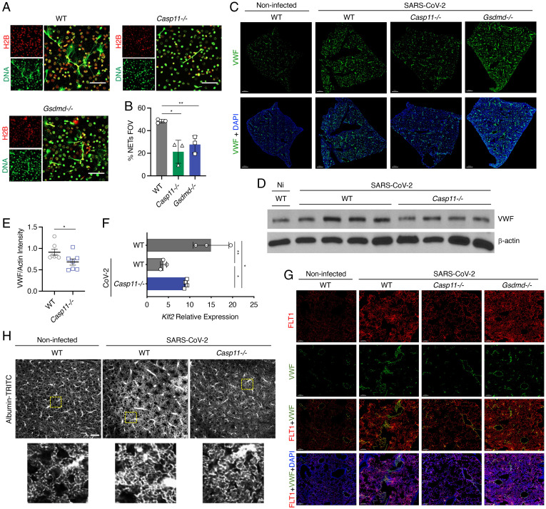
Severe acute respiratory syndrome coronavirus 2 (SARS–CoV-2) is a worldwide health concern, and new treatment strategies are needed. Targeting inflammatory innate immunity pathways holds therapeutic promise, but effective molecular targets remain elusive. Here, we show that human caspase-4 (CASP4) and its mouse homolog, caspase-11 (CASP11), are up-regulated in SARS–CoV-2 infections and that CASP4 expression correlates with severity of SARS–CoV-2 infection in humans. SARS–CoV-2–infected Casp11−/− mice were protected from severe weight loss and lung pathology, including blood vessel damage, compared to wild-type (WT) mice and mice lacking the caspase downstream effector gasdermin-D (Gsdmd−/−). Notably, viral titers were similar regardless of CASP11 knockout. Global transcriptomics of SARS–CoV-2–infected WT, Casp11−/−, and Gsdmd−/− lungs identified restrained expression of inflammatory molecules and altered neutrophil gene signatures in Casp11−/− mice. We confirmed that protein levels of inflammatory mediators interleukin (IL)-1β, IL-6, and CXCL1, as well as neutrophil functions, were reduced in Casp11−/− lungs. Additionally, Casp11−/− lungs accumulated less von Willebrand factor, a marker for endothelial damage, but expressed more Kruppel-Like Factor 2, a transcription factor that maintains vascular integrity. Overall, our results demonstrate that CASP4/11 promotes detrimental SARS–CoV-2–induced inflammation and coagulopathy, largely independently of GSDMD, identifying CASP4/11 as a promising drug target for treatment and prevention of severe COVID-19.

Keywords: SARS–CoV-2; innate immunity; thrombosis.


Conflict of interest statement

The authors declare no competing interest.

Figures

Fig. 1.
Fig. 1.
CASP4 is up-regulated in humans and mice infected with SARS–CoV-2. (A) CASP4 expression levels from RNA sequencing of nasopharyngeal swab samples from patients with no disease, mild SARS–CoV-2, or severe SARS–CoV-2 [GSE145926]; one-way ANOVA with Tukey’s multiple comparisons test. (B) Human lung samples from three donors with healthy lungs or from three donors who died of SARS–CoV-2 were stained for CASP4 (brown). (B, i and ii) Black boxes outline zoomed regions. Scale bars represent 200 μm (B) and 50 μm (i and ii). (C) Quantification of CASP4-positive cells from lungs in B; unpaired t test. (DF) Mice were infected for 4 d with mouse-adapted SARS–CoV-2 (MA10, 105 pfu). (D) Casp11 RNA (green, RNAscope ISH) and DAPI (blue) were visualized (3D intensity projection image) in lung sections using 20x objective, scale bars represent 10 μm. (E) Casp11 RNA levels were quantified in lung samples (n = 3) by qRT-PCR; unpaired t test. (F) CASP11 protein levels in lungs described in D (n = 3) were examined by Western blot. (G) K18-hACE2 mice were infected for 4 d with human SARS–CoV-2 (WA1, 105 pfu), and Casp11 RNA levels were quantitated in lung samples (n = 4) by qRT-PCR; unpaired t test. Error bars in A, C, E, and G represent SD of the mean. *P < 0.05, **P < 0.005, ****P < 0.0001.
Fig. 2.
Fig. 2.
Casp11−/− mice show decreased SARS–CoV-2 infection severity without affecting viral titers but by modulating specific inflammatory programs. (AC) WT, Casp11−/−, and Gsdmd−/− mice were infected with SARS–CoV-2 (MA10, 105 pfu). (A) Weight loss was tracked for 7 d. *P < 0.05, **P < 0.005, ****P < 0.0001; ANOVA with Bonferroni’s multiple comparisons test, day 0 to 4 WT (n = 7), Casp11−/− (n = 10), Gsdmd−/− (n = 9); day 5 to 7 WT (n = 4), Casp11−/− (n = 7), Gsdmd−/− (n = 6). Error bars in represent SEM. (B) TCID50 viral titers were quantified in lung tissue homogenates. Error bars represent SD of the mean. (C) Sections from noninfected control lungs or lungs collected at 4 d after infection were stained for SARS–CoV-2 nucleocapsid protein (brown staining, images representative of at least three mice per group). Insets outline zoomed regions. (D–F) WT, Casp11−/−, and Gsdmd−/− mice (n = 3) were infected with SARS–CoV-2 (MA10, 105 pfu) for 2 d. RNA was extracted from lungs and subjected to RNA sequencing. (D) PCA of SARS–CoV-2–infected lung gene expression with points representing individual WT (gray), Casp11−/−(blue), and Gsdmd−/− (green) mice. PC1 and PC2 represent principal component1 and 2, respectively. (E) Top 30 significant Gene Ontology Biological Pathways are depicted. Node size indicates the number of transcripts within each functional category. Edges connect overlapping gene sets. Numbers represent individual replicates, and color indicates relative up-regulation (red) or down-regulation (blue) in gene expression. TNF: tumor necrosis factor alpha, ROS: reactive oxygen species. (F) Heatmap of significantly changed cytokine and chemokine genes when comparing Casp11−/− infected lungs versus WT. Expression scaling is relative to WT and Gsdmd−/− mice for comparisons (n = 3) (P < 0.05).
Fig. 3.
Fig. 3.
Casp11−/− mice show decreased lung inflammation, less neutrophil recruitment, and altered neutrophil function in response to SARS–CoV-2 infection. (A and B) WT and Casp11−/− mice were infected with SARS–CoV-2 (MA10, 105 pfu). (A) Lung sections from day 4 after infection were stained with H&E to visualize lung damage and airway consolidation. (B) Lung sections as in A were analyzed by the color deconvolution method to quantify cellularity as an indicator of cellular infiltration and alveolar wall thickening; ANOVA with Tukey’s multiple comparisons test. (C and D) Lung homogenates from 2 or 4 d after infection were analyzed by ELISA for detection of CXCL1, IL-1β, or IL-6; ANOVA with Tukey’s multiple comparisons test. (E and F) Macrophages were purified from lungs of mice of the indicated genotype. The cells were infected with mouse-adapted SARS–CoV-2 (multiplicity of infection [MOI] 1 for 24 h). Cell supernatants were analyzed by ELISA, or cellular RNA was analyzed by qRT-PCR for the indicated chemokine/cytokines; ANOVA with Tukey’s multiple comparisons test. (G) Heatmap of significantly changed neutrophil-related genes comparing Casp11−/− infected lungs versus WT (P < 0.05). Expression scaling is relative to WT and Gsdmd−/− mice for comparisons. Numbers represent individual replicates, and color indicates relative up-regulation (red) or down-regulation (blue) in gene expression. (H and I) Lung sections of day 2 SARS–CoV-2–infected WT, Casp11−/−, and Gsdmd−/−mice (n = 5) stained with neutrophil marker Ly6G (H) and quantified in I. (J and K) Flow cytometry of lung single-cell suspensions previously gated on CD45+ cells from WT (n = 4), Casp11−/− (n = 6), and Gsdmd−/− (n = 4) mice (J) as in H and quantified in K. All error bars represent SEM. *P < 0.05, **P < 0.005.
Fig. 4.
Fig. 4.
Casp11−/− neutrophils undergo less NETosis, and Casp11−/− mice show decreased indicators of coagulopathy in lungs after SARS–CoV-2 infection. (A) Neutrophils from WT, Casp11−/−, and Gsdmd−/− mice were treated with PMA, and NET formation was visualized by staining with anti-mouse Histone 2b (H2B) (red) and anti-dsDNA (green). Images were captured at 60x magnification. (B) Percentage of cells undergoing NETosis as averaged from 10 fields of view (FOVs) for each experimental replicate. Error bars represent SEM. (CG) WT, Gsdmd−/−, and Casp11−/− mice were infected with SARS–CoV-2 (MA10, 105 pfu). Lungs were collected at day 4 after infection. (C) RNA for VWF (green) was stained by RNAscope ISH, and nuclei were stained with DAPI (blue). Images were captured by a 20x objective in a 3D stitched panoramic view. Intensity projection images were created using IMARIS software. Scale bars represent 500 μm. (D and E) Western blotting of lung homogenates from noninfected WT and SARS–CoV-2–infected WT and Casp11−/− mice (D) as described in C were quantified in E; unpaired t test. Error bars represent SEM. (F) qRT-PCR quantification of KLF2 in the lungs of mice as described in C; unpaired t test. Error bars represent SEM. (G) Confocal microscopy for the colocalization of VWF RNA (green) with endothelial VEGF receptor subtype 1 (FLT1, red) in the lungs of mice as described in C. Nuclei were stained with DAPI (blue). Images were captured with a 20x objective in a z-stack 3D view and visualized using intensity projection function of IMARIS software. (H) Vasculature imaging of intact lungs 4 d after infection. Below: Higher-magnification view of the regions in yellow boxes. (Scale bar, 200 μm.) *P < 0.05, **P < 0.005.

References

    1. Adil M. T., et al. , SARS-CoV-2 and the pandemic of COVID-19. Postgrad. Med. J. 97, 110–116 (2021). - PMC - PubMed
    1. Das A., Roy S., Swarnakar S., Chatterjee N., Understanding the immunological aspects of SARS-CoV-2 causing COVID-19 pandemic: A therapeutic approach. Clin. Immunol. 231, 108804 (2021). - PMC - PubMed
    1. Lavillegrand J. R., et al. , Elevated plasma IL-6 and CRP levels are associated with adverse clinical outcomes and death in critically ill SARS-CoV-2 patients: Inflammatory response of SARS-CoV-2 patients. Ann. Intensive Care 11, 9 (2021). - PMC - PubMed
    1. Rodríguez C., et al. , Pulmonary endothelial dysfunction and thrombotic complications in patients with COVID-19. Am. J. Respir. Cell Mol. Biol. 64, 407–415 (2021). - PMC - PubMed
    1. Ladikou E. E., et al. , Von Willebrand factor (vWF): Marker of endothelial damage and thrombotic risk in COVID-19? Clin. Med. (Lond.) 20, e178–e182 (2020). - PMC - PubMed

Publication types