. 2022 Aug 10;30(8):1103-1111.e6.
doi: 10.1016/j.chom.2022.04.017. Epub 2022 May 6.

SARS-CoV-2 Omicron sublineages show comparable cell entry but differential neutralization by therapeutic antibodies

Affiliations

SARS-CoV-2 Omicron sublineages show comparable cell entry but differential neutralization by therapeutic antibodies

Prerna Arora et al. Cell Host Microbe. .

Abstract

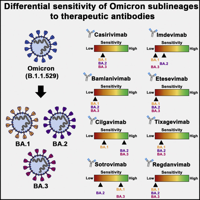
The Omicron variant of SARS-CoV-2 evades antibody-mediated neutralization with unprecedented efficiency. At least three Omicron sublineages have been identified-BA.1, BA.2, and BA.3-and BA.2 exhibits increased transmissibility. However, it is currently unknown whether BA.2 differs from the other sublineages regarding cell entry and antibody-mediated inhibition. Here, we show that BA.1, BA.2, and BA.3 enter and fuse target cells with similar efficiency and in an ACE2-dependent manner. However, BA.2 was not efficiently neutralized by seven of eight antibodies used for COVID-19 therapy, including Sotrovimab, which robustly neutralized BA.1. In contrast, BA.2 and BA.3 (but not BA.1) were appreciably neutralized by Cilgavimab, which could constitute a treatment option. Finally, all sublineages were comparably and efficiently neutralized by antibodies induced by BNT162b2 booster vaccination after previous two-dose homologous or heterologous vaccination. Collectively, the Omicron sublineages show comparable cell entry and neutralization by vaccine-induced antibodies but differ in susceptibility to therapeutic antibodies.

Keywords: ACE2; Omicron; SARS-CoV-2; antibody; neutralization; sotrovimab; spike; vaccine.


Conflict of interest statement

Declaration of interests The authors declare no competing interests.

Figures

None
Graphical abstract
Figure 1
Figure 1
S proteins of Omicron sublineages do not exhibit major differences in ACE2 usage or ability to drive cell-cell and virus-cell fusion (A) Schematic overview of the SARS-CoV-2 spike (S) protein domain structure (left) and summary of the mutations found in the different Omicron sublineages (right; numbering is according to the S protein of SARS-CoV-2 B.1). S protein residues that are identical between the S proteins of some Omicron sublineages and B.1 are marked in green, whereas mutated residues are highlighted in red (note: the BA.1 S protein harbors an insertion between amino acid residues 214 and 215). Further, mutations found in all Omicron sublineages are indicated by a circle. Abbreviations: NTD, N-terminal domain; RBD, receptor-binding domain; TD, transmembrane domain; S1/S2 and S2’, cleavage sites in the S protein. (B) S-protein-driven cell entry. We added particles bearing the indicated S proteins (or no S protein) to the indicated cell lines and analyzed cell entry by measuring the activity of virus-encoded firefly luciferase in cell lysates 16–18 h after inoculation. Presented are the average (mean) data from 6–12 biological replicates (each conducted with four technical replicates) in which cell entry was normalized against B.1 (set as 1). Error bars show the SEM. Statistical significance was assessed by two-tailed Student’s t tests (p > 0.05, not significant [ns]; p ≤ 0.05, ; p ≤ 0.01, ∗∗; p ≤ 0.001, ∗∗∗). Please also see Figure S1. (C) ACE2 binding efficiency. 293T cells transiently expressing the indicated S proteins (or no S protein) where first incubated with soluble ACE2 fused to the Fc portion of human immunoglobulin G (solACE2-Fc) and subsequently incubated with an Fc-specific AlexaFluor-488-coupled secondary antibody; then, solACE2-Fc binding was analyzed by flow cytometry. Presented are the average (mean) data from six biological replicates (each conducted with single samples) in which ACE2 binding was normalized against B.1 (set as 1). Error bars show the SEM. Statistical significance was assessed by two-tailed Student’s t tests (p > 0.05, not significant [ns]; p ≤ 0.05, ; p ≤ 0.01, ∗∗). (D) Blockade of S-protein-driven cell entry by an anti-ACE2 antibody. Vero cells were preincubated with serial dilutions of anti-ACE2 antibody before particles bearing the indicated S proteins were added. S-protein-driven cell entry was analyzed and normalized to samples without anti-ACE2 antibody (set as 1). Presented are the average (mean) data of three biological replicates, each performed with four technical replicates. Error bars show the SEM. Statistical significance was assessed by two-way analysis of variance with Dunnett’s post hoc tests (p > 0.05, not significant [ns]; p ≤ 0.05, ; p ≤ 0.01, ∗∗; p ≤ 0.001, ∗∗∗). Please also see Tables S1 and S2. (E) Blockade of S-protein-driven cell entry by solACE2-Fc. Particles bearing the indicated S proteins were preincubated with serial dilutions of solACE2-Fc (or no solACE2-Fc) before being added to Vero cells. S-protein-driven cell entry was analyzed and normalized to samples without solACE2-Fc (= 0% inhibition). Presented are the average (mean) data of three biological replicates, each performed with four technical replicates. Error bars show the SEM. Statistical significance was assessed by two-way analysis of variance with Dunnett’s post hoc tests (p > 0.05, not significant [ns]). (F) Usage of mouse and bat ACE2 by Omicron S proteins. BHK-21 cells transiently expressing human, mouse, or horseshoe bat (Rhinolophus pearsonii) ACE2 orthologs (or no ACE2) were inoculated with particles bearing the indicated S proteins or VSV glycoprotein (VSV-G). Cell entry of pseudovirus particles was analyzed and normalized to particles bearing no viral glycoprotein (set as 1; indicated by dashed line). Presented are the average (mean) data of three biological replicates, each performed with four technical replicates. Error bars show the SEM. Statistical significance was assessed by two-tailed Student’s t tests (p > 0.05, not significant [ns]; p ≤ 0.05, ). (G) Qualitative fusion assay. A549-ACE2 cells transfected to express the indicated S proteins (or no S protein) were fixed 24 h after transfection and stained with May-Gruenwald and Giemsa solution before microscopic images were taken (scale bar, 500 μm). Arrowheads indicate small syncytia in cells expressing the S proteins of the different Omicron sublineages. (H) Quantitative fusion assay. 24 h after transfection, 293T cells transiently expressing the indicated S proteins (or no S protein) along with the beta-galactosidase alpha fragment were resuspended and seeded on top of A549-ACE2 cells transiently expressing the beta-galactosidase omega fragment. After an additional 24 h of incubation, beta-galactosidase substrate was added, and luminescence was recorded. Presented are the average (mean) data of three biological replicates, each performed with four technical replicates. Error bars show the SEM. Statistical significance was assessed by two-tailed Student’s t tests (p ≤ 0.05, ).
Figure 2
Figure 2
S proteins of Omicron sublineages differ in terms of their resistance to therapeutic antibodies (A) Schematic overview of the SARS-CoV-2 S protein RBD (numbering is according to the S protein of SARS-CoV-2 B.1). Mutations found in the RBDs of the Omicron sublineages (compared with the B.1 S protein) are highlighted in red, whereas identical amino acid residues are marked in green. RBD residues that make direct contact with ACE2 or that are recognized as epitopes for therapeutic antibodies are highlighted. (B) Particles bearing the indicated S proteins were preincubated with serial dilutions of individual monoclonal antibodies used for COVID-19 treatment or cocktails thereof or an irrelevant control antibody (hIgG) before being added to Vero cells. Of note, for antibody cocktails, we used each antibody at half concentration in order to keep total antibody concentrations constant. S-protein-driven cell entry was analyzed and normalized to samples without antibody (= 0% inhibition). Presented are the average (mean) data of three biological replicates, each performed with four technical replicates. Error bars show the SEM. Statistical significance was assessed by two-way analysis of variance with Dunnett’s post hoc tests (p > 0.05, not significant [ns]; p ≤ 0.05, ; p ≤ 0.01, ∗∗; p ≤ 0.001, ∗∗∗). Please see also Tables S1 and S2.
Figure 3
Figure 3
S proteins of Omicron sublineages do not differ in sensitivity to neutralization by antibodies elicited upon triple vaccination with BNT162b2 or BNT162b2/ChAdOx1-S (A) Left: particles bearing the indicated S proteins were preincubated with serial dilutions of serum from individuals vaccinated three times with BNT162b2 (BNT) before being added to Vero cells. S-protein-driven cell entry was analyzed and used for calculating the neutralizing titer 50 (NT50). Presented are the combined data for 15 sera (black lines show the geometric mean; the dashed line indicates the lowest serum dilution tested). Numerical values above the graph indicate the proportion of sera with reactivity against the respective S-protein-bearing particles and the geometric mean NT50. The statistical significance of differences between individual groups was assessed by two-tailed Mann-Whitney test (p > 0.05, not significant [ns]; p ≤ 0.05, ; p ≤ 0.01, ∗∗; p ≤ 0.001, ∗∗∗). Right: individual NT50 values per serum (ranked according to neutralizing activity against B.1) and fold change in neutralizing activity compared with that of B.1. Please also see Figure S2 and Table S3. (B) The experiment was performed as described for (A), but this time 15 sera from individuals who had been vaccinated twice with ChAdOx1-S (AZ) and then once with BNT162b2 (BNT) were analyzed. Please also see Figure S2 and Table S3. (C) The experiment was performed as described for (A), but this time 15 sera from individuals who had been vaccinated once with ChAdOx1-S (AZ) and then twice with BNT162b2 (BNT) were analyzed. Please also see Figure S2 and Table S3.

References

    1. Abu-Raddad L.J., Chemaitelly H., Ayoub H.H., AlMukdad S., Yassine H.M., Al-Khatib H.A., Smatti M.K., Tang P., Hasan M.R., Coyle P., et al. Effect of mRNA vaccine boosters against SARS-CoV-2 omicron infection in Qatar. N. Engl. J. Med. 2022 doi: 10.1056/NEJMoa2200797. NEJMoa2200797. - DOI - PMC - PubMed
    1. Accorsi E.K., Britton A., Fleming-Dutra K.E., Smith Z.R., Shang N., Derado G., Miller J., Schrag S.J., Verani J.R. Association between 3 doses of mRNA COVID-19 vaccine and symptomatic infection caused by the SARS-CoV-2 omicron and Delta variants. JAMA. 2022;327:639–651. doi: 10.1001/jama.2022.0470. - DOI - PMC - PubMed
    1. Altarawneh H.N., Chemaitelly H., Hasan M.R., Ayoub H.H., Qassim S., AlMukdad S., Coyle P., Yassine H.M., Al-Khatib H.A., Benslimane F.M., et al. Protection against the omicron variant from previous SARS-CoV-2 infection. N. Engl. J. Med. 2022;386:1288–1290. doi: 10.1056/NEJMc2200133. - DOI - PMC - PubMed
    1. Arora P., Sidarovich A., Kruger N., Kempf A., Nehlmeier I., Graichen L., Moldenhauer A.S., Winkler M.S., Schulz S., Jack H.M., et al. B.1.617.2 enters and fuses lung cells with increased efficiency and evades antibodies induced by infection and vaccination. Cell Rep. 2021;37:109825. doi: 10.1016/j.celrep.2021.109825. - DOI - PMC - PubMed
    1. Arora P., Zhang L., Rocha C., Sidarovich A., Kempf A., Schulz S., Cossmann A., Manger B., Baier E., Tampe B., et al. Comparable neutralisation evasion of SARS-CoV-2 omicron subvariants BA.1, BA.2, and BA.3. Lancet Infect. Dis. 2022 doi: 10.1016/s1473-3099(22)00224-9(22)00224-9. S1473-3099(22)00224-9. - DOI - PMC - PubMed

Supplementary concepts

LinkOut - more resources