

Conserved circuits for direction selectivity in the primate retina
- PMID: 35588744
- PMCID: PMC9205626
- DOI: 10.1016/j.cub.2022.04.056
Conserved circuits for direction selectivity in the primate retina
Abstract
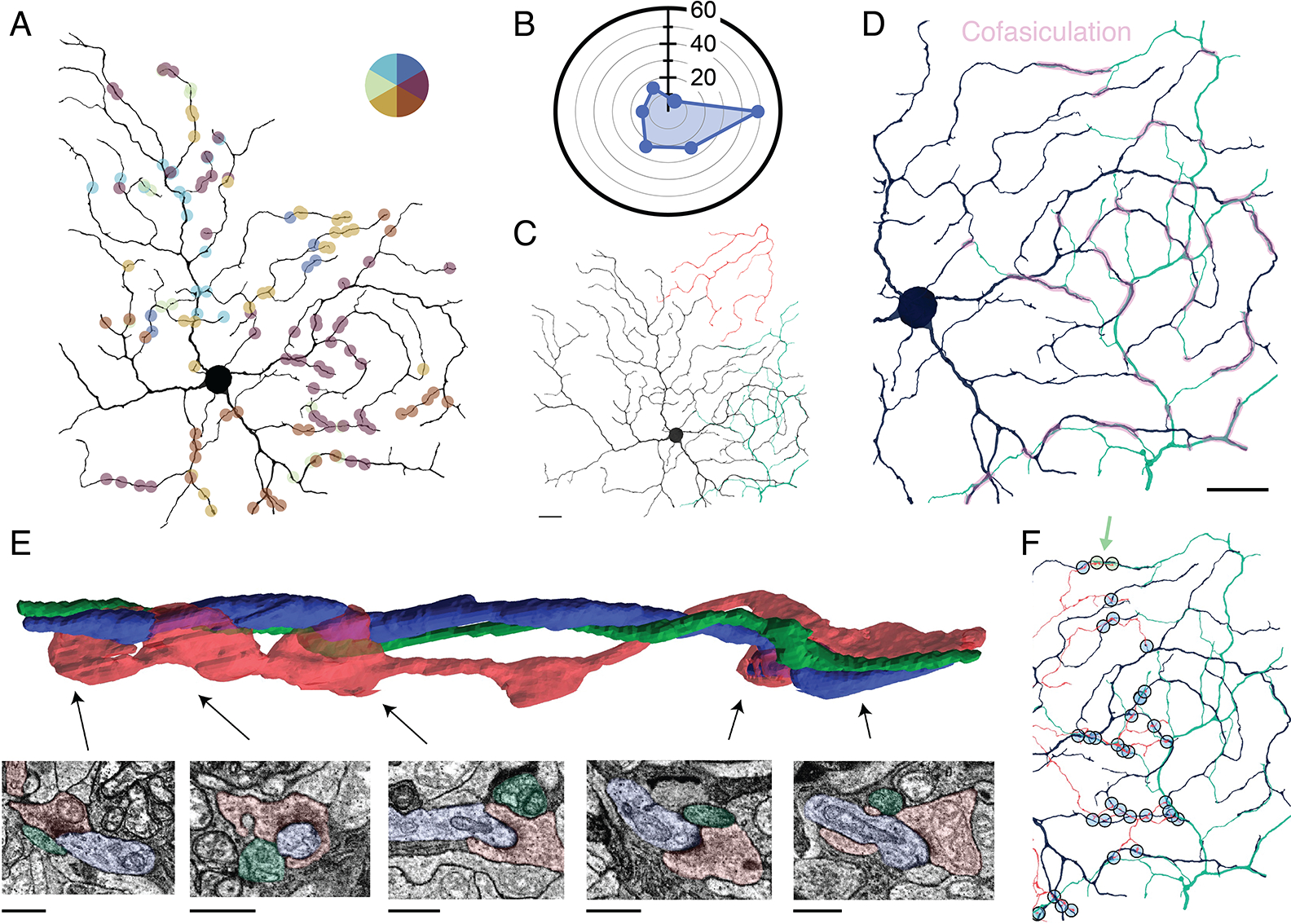
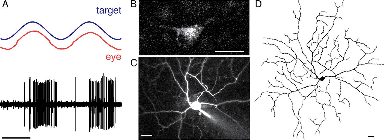
The detection of motion direction is a fundamental visual function and a classic model for neural computation. In the non-primate retina, direction selectivity arises in starburst amacrine cell (SAC) dendrites, which provide selective inhibition to direction-selective retinal ganglion cells (dsRGCs). Although SACs are present in primates, their connectivity and the existence of dsRGCs remain open questions. Here, we present a connectomic reconstruction of the primate ON SAC circuit from a serial electron microscopy volume of the macaque central retina. We show that the structural basis for the SACs' ability to confer directional selectivity on postsynaptic neurons is conserved. SACs selectively target a candidate homolog to the mammalian ON-sustained dsRGCs that project to the accessory optic system (AOS) and contribute to gaze-stabilizing reflexes. These results indicate that the capacity to compute motion direction is present in the retina, which is earlier in the primate visual system than classically thought.
Keywords: accessory optic system; bipolar cells; connectomics; direction selectivity; motion processing; primate; retina; retinal circuitry; retinal ganglion cells; starburst amacrine cells.
Copyright © 2022 The Author(s). Published by Elsevier Inc. All rights reserved.
Conflict of interest statement
Declaration of interests The authors declare no competing interests.
Figures

Similar articles
-
Origins of direction selectivity in the primate retina.Nat Commun. 2022 May 23;13(1):2862. doi: 10.1038/s41467-022-30405-5. Nat Commun. 2022. PMID: 35606344 Free PMC article.
-
A structural basis for omnidirectional connections between starburst amacrine cells and directionally selective ganglion cells in rabbit retina, with associated bipolar cells.Vis Neurosci. 2002 Mar-Apr;19(2):145-62. doi: 10.1017/s0952523802191139. Vis Neurosci. 2002. PMID: 12385627
-
Species-specific wiring for direction selectivity in the mammalian retina.Nature. 2016 Jul 7;535(7610):105-10. doi: 10.1038/nature18609. Epub 2016 Jun 22. Nature. 2016. PMID: 27350241 Free PMC article.
-
Neural Mechanisms of Motion Processing in the Mammalian Retina.Annu Rev Vis Sci. 2018 Sep 15;4:165-192. doi: 10.1146/annurev-vision-091517-034048. Epub 2018 Aug 10. Annu Rev Vis Sci. 2018. PMID: 30095374 Review.
-
Direction selectivity in the retina.Curr Opin Neurobiol. 2002 Aug;12(4):405-10. doi: 10.1016/s0959-4388(02)00337-9. Curr Opin Neurobiol. 2002. PMID: 12139988 Review.
Cited by
-
Primate eye tracking with carbon-nanotube-paper-composite based capacitive sensors and machine learning algorithms.J Neurosci Methods. 2024 Oct;410:110249. doi: 10.1016/j.jneumeth.2024.110249. Epub 2024 Aug 14. J Neurosci Methods. 2024. PMID: 39151657
-
Synaptic Origins of the Complex Receptive Field Structure in Primate Smooth Monostratified Retinal Ganglion Cells.eNeuro. 2024 Jan 30;11(1):ENEURO.0280-23.2023. doi: 10.1523/ENEURO.0280-23.2023. Print 2024 Jan. eNeuro. 2024. PMID: 38290840 Free PMC article.
-
Target identification and validation of the alpha7 nicotinic acetylcholine receptor as a potential therapeutic target in retinal disease.Front Ophthalmol (Lausanne). 2023 Jul 24;3:1190439. doi: 10.3389/fopht.2023.1190439. eCollection 2023. Front Ophthalmol (Lausanne). 2023. PMID: 38983049 Free PMC article.
-
Spatiotemporal properties of glutamate input support direction selectivity in the dendrites of retinal starburst amacrine cells.Elife. 2022 Nov 8;11:e81533. doi: 10.7554/eLife.81533. Elife. 2022. PMID: 36346388 Free PMC article.
-
Rejection of inappropriate synaptic partners in mouse retina mediated by transcellular FLRT2-UNC5 signaling.Dev Cell. 2023 Oct 23;58(20):2080-2096.e7. doi: 10.1016/j.devcel.2023.07.011. Epub 2023 Aug 8. Dev Cell. 2023. PMID: 37557174 Free PMC article.
References
-
- Borst A, and Helmstaedter M (2015). Common circuit design in fly and mammalian motion vision. Nat. Neurosci. 18, 1067–1076. - PubMed
-
- Pasternak T, and Tadin D (2020). Linking neuronal direction selectivity to perceptual decisions about visual motion. Annu. Rev. Vis. Sci. 6, 335–362. - PubMed
-
- Borst A, and Euler T (2011). Seeing things in motion: models, circuits, and mechanisms. Neuron 71, 974–994. - PubMed
Publication types
MeSH terms
Grants and funding
LinkOut - more resources
Full Text Sources
Miscellaneous
