. 2022 May 19;13(1):2770.
doi: 10.1038/s41467-022-30430-4.

Structural insights into the mechanism of pancreatic KATP channel regulation by nucleotides

Affiliations

Structural insights into the mechanism of pancreatic KATP channel regulation by nucleotides

Mengmeng Wang et al. Nat Commun. .

Abstract

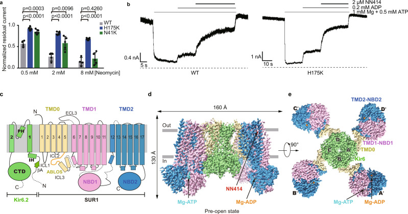
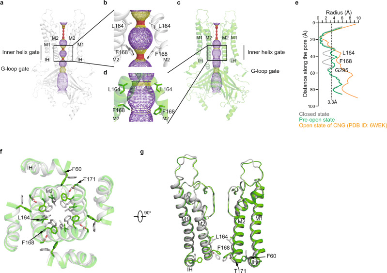
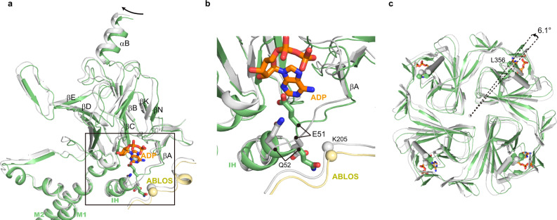
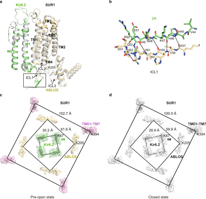
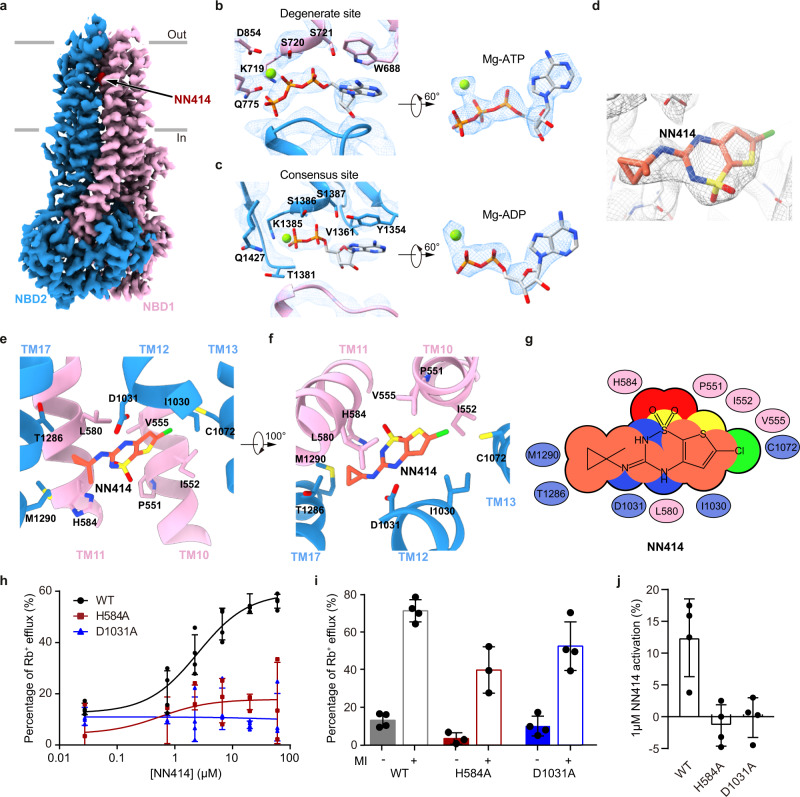
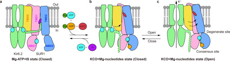
ATP-sensitive potassium channels (KATP) are metabolic sensors that convert the intracellular ATP/ADP ratio to the excitability of cells. They are involved in many physiological processes and implicated in several human diseases. Here we present the cryo-EM structures of the pancreatic KATP channel in both the closed state and the pre-open state, resolved in the same sample. We observe the binding of nucleotides at the inhibitory sites of the Kir6.2 channel in the closed but not in the pre-open state. Structural comparisons reveal the mechanism for ATP inhibition and Mg-ADP activation, two fundamental properties of KATP channels. Moreover, the structures also uncover the activation mechanism of diazoxide-type KATP openers.


Conflict of interest statement

The authors declare no competing interests.

Figures

Fig. 1
Fig. 1. Structure of the pancreatic KATP channel (H175Kcryo-EM) in the pre-open state.
a Neomycin inhibition of the inside-out currents of the KATP channel. Wild type (WT), H175K, and N41K mutants of Kir6.2 were co-expressed with wild type SUR1 for recordings. Data are shown as mean ± SD. WT n = 4, H175K n = 6, N41K n = 5 independent patches, respectively. p values were calculated by unpaired two-tailed t-test and were indicated above. b Representative inside-out recordings of KATP channel formed by the wild-type Kir6.2 or the H175K mutant. c Topology of Kir6.2 and SUR1 subunits. PH, pore helix; ECL, extracellular loop; ICL, intracellular loop; IH, interfacial helix; CTD, cytoplasmic domain; TMD, transmembrane domain; NBD, nucleotide-binding domain. Transmembrane helices are shown as cylinders. The phospholipid bilayer is shown as thick gray lines. Kir6.2, SUR1 TMD0-ICL3 fragment, TMD1-NBD1, and TMD2-NBD2 are shown in green, yellow, violet, and blue, respectively. d Side view of the KATP complex in the pre-open state. Mg-ADP, Mg-ATP, and NN414 are shown in orange, cyan, and red, respectively. e Bottom view of the KATP channel in the pre-open state from the intracellular side. Source data are provided as a Source Data file.
Fig. 2
Fig. 2. The conformational changes of Kir6.2 TMD during KATP channel opening.
a Side view of Kir6.2 subunits of H175Kcryo-EM in the closed state. The ion conduction pathway along the pore is shown as dots and colored as red, yellow, and purple according to the pore radii of <1.4, 1.4–3.3, and >3.3 Å. M1, M2, and IH are labeled. For clarity, the subunits in front and in the back were omitted. b Close-up view of M2 helices in (a) with gate residues shown as sticks (L164 and F168). c Side view of Kir6.2 subunits of H175Kcryo-EM in the pre-open state. d Close-up view of M2 helices in (c). e Calculated pore profiles of the H175Kcryo-EM closed state (gray), H175Kcryo-EM pre-open state (green), and the open state of CNG channel (PDB ID: 6WEK) (orange). The size of a fully hydrated potassium ion (3.3 Å) is shown as dashes. f Structural comparison of the transmembrane domain between the closed state (gray) and the pre-open state (green). g A 90° rotated view of (d).
Fig. 3
Fig. 3. Conformational changes of Kir6.2 CTD during KATP channel opening.
a Conformational changes of the nucleotide-binding site between the closed (gray) and the pre-open (green) states of H175Kcryo-EM. The ADP bound in the closed state is shown as sticks in orange. b Close-up view of the nucleotide-binding site boxed in (a). c Bottom view of the Kir6.2 CTD. The rotation angle between CTDs was measured using Cα positions of L356 of Kir6.2 as marker atoms (shown as spheres).
Fig. 4
Fig. 4. Conformational changes of SUR1 during KATP channel opening.
a Structural comparison of SUR1 TMD0 between the closed state (gray) and the pre-open state (colored) of H175Kcryo-EM. b Close-up view of the interaction between βA of Kir6.2 and SUR1-ICL1 boxed in (a). Putative hydrogen bondings are shown as dashes. c, d Bottom view of the structural arrangement of the KATP complex during channel opening. Cα positions of K67 on IH of Kir6.2, K205 on ABLOS of SUR1, and K397 on M7 of SUR1 are shown as spheres. Distances of marker atoms in the pre-open state (colored) and the closed state (gray) are shown in (c, d), respectively.
Fig. 5
Fig. 5. Structure of the SUR1 subunit in complex with NN414 and Mg-nucleotides.
a Cryo-EM density map of SUR1 in complex with Mg-nucleotides and NN414, viewed from the side. The approximate position of the lipid bilayer is indicated by gray bars. TMD1-NBD1, TMD2-NBD2, and NN414 are colored in pink, blue and red, respectively. For better visualization of the position of NN414, a fragment of TMD2 in front of NN414 was omitted. b Close-up views of electron densities at the degenerate site. NBD1, NBD2, nucleotides, and Mg2+ are colored in pink, blue, gray, and green, respectively. c Electron densities at the consensus sites. d NN414 density (orange) in the SUR1 subunit (gray). The map is shown as mesh and the protein is shown as sticks. e, f Close-up views of the NN414-binding site. TMD1 and TMD2 are colored in pink and blue, respectively. NN414 (orange) and residues that interact with NN414 are shown as sticks. g Cartoon representation of the interaction between NN414 and SUR1. The key residues on TMD1 and TMD2 are shown as pink and blue ovals, respectively. h The dose-response activation curves of SUR1- Kir6.2 KATP channel by NN414 measured by Rb+ efflux assay. Curves were fitted to the Hill equation. Data are shown as mean ± SD and WT and D1031A n = 4, H584A n = 3 independent Rb+ efflux assays, respectively. i Effects of metabolism inhibitors (MI) on KATP channel containing various SUR1 mutants. Data are shown as mean ± SD and WT and D1031A n = 4, H584A n = 3 independent Rb+ efflux assays corresponding to (h). j KATP channel activation by 1 µM NN414 in the presence of 0.1 mM Mg-ATP. Data are shown as mean ± SD and n = 3 independent patches. Source data are provided as a Source Data file.
Fig. 6
Fig. 6. Model for the KATP channel activation by Mg-nucleotides and KATP opener.
ac Side view of the cartoon model of the KATP channel. For simplicity, a pair of Kir6 subunits and one SUR2 subunit are shown. Kir6, TMD0, TMD1-NBD1, TMD2-NBD2, Mg2+, ATP, ADP, insulin secretagogue (IS), and KATP opener (KCO) are colored in green, yellow, pink, blue, dark green, cyan, orange, purple, and red, respectively. The flexible KNtp in the Mg-nucleotide and KATP opener-bound state is outlined by dashed lines.

Similar articles

Cited by

References

    1. Nichols CG. KATP channels as molecular sensors of cellular metabolism. Nature. 2006;440:470–476. doi: 10.1038/nature04711. - DOI - PubMed
    1. Ashcroft FM. K(ATP) channels and insulin secretion: a key role in health and disease. Biochem. Soc. Trans. 2006;34:243–246. doi: 10.1042/BST0340243. - DOI - PubMed
    1. Vishwakarma VK, et al. Mechanistic Pathways of ATP Sensitive Potassium Channels Referring to Cardio-Protective Effects and Cellular Functions. Drug Res. (Stuttg.) 2019;69:365–373. doi: 10.1055/a-0806-7207. - DOI - PubMed
    1. Brayden JE. Functional roles of KATP channels in vascular smooth muscle. Clin. Exp. Pharmacol. Physiol. 2002;29:312–316. doi: 10.1046/j.1440-1681.2002.03650.x. - DOI - PubMed
    1. Pipatpolkai T, Usher S, Stansfeld PJ, Ashcroft FM. New insights into KATP channel gene mutations and neonatal diabetes mellitus. Nat. Rev. Endocrinol. 2020;16:378–393. doi: 10.1038/s41574-020-0351-y. - DOI - PubMed

Publication types

MeSH terms