. 2022 May 19;12(1):8382.
doi: 10.1038/s41598-022-12031-9.

Microfluidic-assisted synthesis and modeling of stimuli-responsive monodispersed chitosan microgels for drug delivery applications

Affiliations

Microfluidic-assisted synthesis and modeling of stimuli-responsive monodispersed chitosan microgels for drug delivery applications

Omid Sartipzadeh et al. Sci Rep. .

Abstract

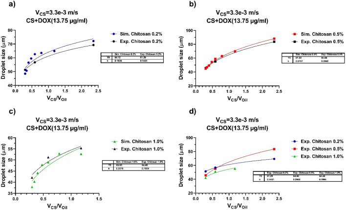
Droplet microfluidic has been established to synthesize and functionalize micro/nanoparticles for drug delivery and screening, biosensing, cell/tissue engineering, lab-on-a-chip, and organ-on-a-chip have attracted much attention in chemical and biomedical engineering. Chitosan (CS) has been suggested for different biomedical applications due to its unique characteristics, such as antibacterial bioactivities, immune-enhancing influences, and anticancer bioactivities. The simulation results exhibited an alternative for attaining visions in this complex method. In this regard, the role of the flow rate ratio on the CS droplet features, including the generation rate and droplet size, were thoroughly described. Based on the results, an appropriate protocol was advanced for controlling the CS droplet properties for comparing their properties, such as the rate and size of the CS droplets in the microchip. Also, a level set (LS) laminar two-phase flow system was utilized to study the CS droplet-breaking process in the Flow Focused-based microchip. The outcomes demonstrated that different sizes and geometries of CS droplets could be established via varying the several parameters that validated addressing the different challenges for several purposes like drug delivery (the droplets with smaller sizes), tissue engineering, and cell encapsulation (the droplets with larger sizes), lab-on-a-chip, organ-on-a-chip, biosensing and bioimaging (the droplets with different sizes). An experimental study was added to confirm the simulation results. A drug delivery application was established to verify the claim.


Conflict of interest statement

The authors declare no competing interests.

Figures

Figure 1
Figure 1
The pattern of the MFFD applied in simulations: meshes and boundaries situations specified for the microfluidic droplet generatrix in the two-dimensions model.
Figure 2
Figure 2
The microscopy and two-dimensional images of mechanism formation of CS + DOX microgel droplet injunction, (a) lag stage, (b) filling stage, (c) necking stage, (d) detachment. The CS + DOX and vegetable oil phase velocities were adjusted at 3.3 and 11.1 mm/s. The Concentration of CS and DOX are 0.2% and 13.75 (µg/ml), respectively.
Figure 3
Figure 3
The CS with a concentration of 0.2% and 13.75 mg of DOX per ml of CS solution. The scanning electron microscope (SEM) of outcome CS + DOX microgel droplet of experimental result that the volumetric flow rate of CS + DOX phase was 3.3 mm/s and the volumetric flow rate of oil phase was 11.1 mm/s.
Figure 4
Figure 4
The procedure of the pressure slope at the P point for the duration of droplet configuration. The P point decides the point situated at the main duct entry. It reverberates the development of the droplet formation process. Three steps formation of droplet: (I) Lag, (II) Filling, and (III) Necking. The CS + DOX velocity is stable at 3.3 mm/s, and the velocity of vegetable oil is equal to 11.1 mm/s.
Figure 5
Figure 5
The computer simulation results in the pressure gradient at the P point. Impact of velocities and concentrations of the CS-DOX upon the droplet formation. The velocities of vegetable oil are changeable at 9.7 and 11.1 mm/s, and the CS-DOX velocities are regulated at 3.3 and 4.2 mm/s. The concentration of CS is (a–c) 0.2%, (d–f) 0.5%, (g–i) 1.0% (w/v), and also the concentration of DOX is 1.375% (w/v).
Figure 6
Figure 6
The dependence of the droplet size produced for each of the CS + DOX concentration to the variations of the velocity ratio of the dispersed phase to the continuous phase. The flow rate of vegetable oil and CS + DOX were selected at the limited area of 1.4 to 11.1 and 3.3 to 9.2 mm/s, respectively. The concentrations of CS are 0.2%, 0.5%, and 1.0% (w/v), and also the concentration of DOX is 1.375% (w/v). For each of subfigure, the CS + DOX velocity is fixed and the flow rate of the vegetable oli varies from 1.4 to 11.1 mm/s. The CS + DOX flow rates are (a) 3.3 mm/s, (b) 4.2 mm/s, (c) 5.0 mm/s, (d) 5.8 mm/s, (e) 6.7 mm/s, (f) 7.5 mm/s, (g) 8.3 mm/s and (h) 9.2 mm/s. As the CS + DOX flow rate increases, some points disappear for each of the three CS + DOX concentrations, indicating that no droplets are formed at this concentration and velocity ratio of the dispersed phase to the continuous phase. Continuous lines are curves plotted on experimental data using the power equation (Y = Y0 * Xk). The values of Y0 and K of each of these curves are shown in the table in each subfigure.
Figure 7
Figure 7
The consequence of velocity and concentration variations of CS-DOX and vegetable oil upon the number of droplet generation per unit time. The flow rate of vegetable oil and CS + DOX were selected at the limited area of 1.4 to 11.1 and 3.3 to 9.2 mm/s, respectively. The concentrations of CS are 0.2%, 0.5% and 1.0% (w/v), and also the concentration of DOX is 1.375% (w/v). For each subfigure, the CS + DOX velocity is fixed and the flow rate of the vegetable oli varies from 1.4 to 11.1 mm/s. The CS + DOX flow rate is (a) 3.3 mm/s, (b) 4.2 mm/s, (c) 5.0 mm/s, (d) 5.8 mm/s, (e) 6.7 mm/s, (f) 7.5 mm/s, (g) 8.3 mm/s and (h) 9.2 mm/s. Continuous lines are curves plotted on experimental data using the power equation (Y = Y0 * Xk). The values of Y0 and K of each of these curves are shown in the table in each subfigure.
Figure 8
Figure 8
Comparisons the size of droplets generated using the computer simulator and experimental experiments. (a) CS 0.2%, (b) CS 0.5%, (c) CS 1.0%, (d) comparison of experimental data of droplet production for three concentrations of CS + DOX at the fixed velocity of the dispersed phase (3.3 mm/s), and velocity ratios of 2.36, 0.59 and 0.27, respectively. DOX concentration is 13.75 (µg/ml).
Figure 9
Figure 9
(a) Standard curve of DOX in ISO buffer at the wavelength of 480 nm. (b) Standard curve of DOX in PBS buffer at the wavelength of 480 nm. (c) Comparing the effect of the free form of DOX on MCF-7 breast cancer cell line in various concentrations. (d) The effect of blank chitosan particles at various diluents on HFF cells. e) Release profile kinetic of DOX from CS particles at various temperatures at pH  7.4. (f) Release kinetics of DOX from CS particles at various temperatures at pH  4.5. (g) Comparing the effect of the free form of DOX and CS-DOX on MCF-7 breast cancer cells. ns no significant difference. ***: P-value < 0.05.
Figure 10
Figure 10
(a) Cellular uptake behavior of DOX by MCF-7 breast cancer cell line. (b) Cellular uptake behavior of CS-DOX by MCF-7 breast cancer cell line. DAPI is used to stain nuclei of cells. Due to its fluorescent nature, DOX causes the cell cytoplasm to turn red on the fluorescent imaging. The images confirm the uptake of the drug by the cells.

Similar articles

Cited by

References

    1. Poonoosamy J, et al. A lab-on-a-chip approach integrating in-situ characterization and reactive transport modelling diagnostics to unravel (Ba, Sr)SO4 oscillatory zoning. Sci. Rep. 2021;11:23678. doi: 10.1038/s41598-021-02840-9. - DOI - PMC - PubMed
    1. Brown CR, et al. Leakage pressures for gasketless superhydrophobic fluid interconnects for modular lab-on-a-chip systems. Microsyst. Nanoeng. 2021;7:69. doi: 10.1038/s41378-021-00287-6. - DOI - PMC - PubMed
    1. Uda MNA, et al. Silica and graphene mediate arsenic detection in mature rice grain by a newly patterned current–volt aptasensor. Sci. Rep. 2021;11:14688. doi: 10.1038/s41598-021-94145-0. - DOI - PMC - PubMed
    1. Subramani IG, et al. 1,1'-Carbonyldiimidazole-copper nanoflower enhanced collapsible laser scribed graphene engraved microgap capacitive aptasensor for the detection of milk allergen. Sci. Rep. 2021;11:20825. doi: 10.1038/s41598-021-00057-4. - DOI - PMC - PubMed
    1. Rahimzadeh Z, et al. A rapid nanobiosensing platform based on herceptin-conjugated graphene for ultrasensitive detection of circulating tumor cells in early breast cancer. Nanotechnol. Rev. 2021;10:744–753. doi: 10.1515/ntrev-2021-0049. - DOI