. 2022 May 19;12(1):8464.
doi: 10.1038/s41598-022-11235-3.

Label-free complete absorption microscopy using second generation photoacoustic remote sensing

Affiliations

Label-free complete absorption microscopy using second generation photoacoustic remote sensing

Benjamin R Ecclestone et al. Sci Rep. .

Abstract

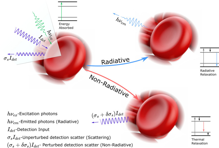
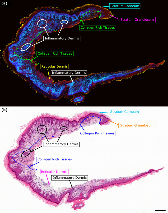
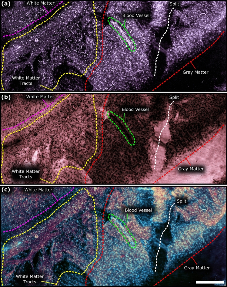
In the past decades, absorption modalities have emerged as powerful tools for label-free functional and structural imaging of cells and tissues. Many biomolecules present unique absorption spectra providing chromophore-specific information on properties such as chemical bonding, and sample composition. As chromophores absorb photons the absorbed energy is emitted as photons (radiative relaxation) or converted to heat and under specific conditions pressure (non-radiative relaxation). Modalities like fluorescence microscopy may capture radiative relaxation to provide contrast, while modalities like photoacoustic microscopy may leverage non-radiative heat and pressures. Here we show an all-optical non-contact total-absorption photoacoustic remote sensing (TA-PARS) microscope, which can capture both radiative and non-radiative absorption effects in a single acquisition. The TA-PARS yields an absorption metric proposed as the quantum efficiency ratio (QER), which visualizes a biomolecule's proportional radiative and non-radiative absorption response. The TA-PARS provides label-free visualization of a range of biomolecules enabling convincing analogues to traditional histochemical staining of tissues, effectively providing label-free Hematoxylin and Eosin (H&E)-like visualizations. These findings establish an effective all-optical non-contact total-absorption microscope for label-free inspection of biological materials.


Conflict of interest statement

Authors Benjamin Ecclestone, Kevan Bell, Deepak Dinakaran, John R. Mackey, and Parsin Haji Reza, all have financial interests in IllumiSonics which has provided funding to the PhotoMedicine Labs. Author Sarah Sparkes does not have any competing interests.

Figures

Figure 1
Figure 1
TA-PARS contrast mechanisms. Each excitation always generates some fraction of radiative and non-radiative relaxation effects. The non-radiative relaxation leads to heat and pressure induced modulations, which in turn cause back-reflected intensity variations in the detection beam. PARS signals are denoted as some change in reflectivity multiplied by the incident detection ((σs+δσs)Idet). The radiative absorption pathway captures optical emissions attributed to radiative relaxation such as stimulated Raman scattering, fluorescence, multiphoton fluorescence, etc. Emissions are denoted as some wavelength and energy optical emission (hνem). The local scattering contrast is captured as the unmodulated backscatter (pre-excitation pulse) of the detection beam. The scattering contrast is denoted as the unperturbed scattering profile multiplied by the incident detection power (σsIdet).
Figure 2
Figure 2
Simplified experimental system setups (i) 515 nm excitation system (b) 266 nm excitation system. (ac) Sample configuration for imaging each type of tissue. Component labels are as follows: HWP half wave plate, LBO lithium triborate crystal, DM dichroic mirror, AD air spaced doublet, PH pinhole, VBE variable beam expander, Col. collimator, Circ. circulator, SF spectral filter, Cond. condenser lens, PD photodiode, 90:10 10:90 splitter, M mirror.
Figure 3
Figure 3
Comparison of the three different contrasts (non-radiative absorption, radiative absorption, and scattering) provided by the TA-PARS system, in a thin section of preserved human brain tissues. (a) 266 nm non-radiative absorption contrast. (b) 266 nm radiative absorption contrast. (c) 405 nm scattering contrast. Scale Bar: 1 mm.
Figure 4
Figure 4
Total-absorption second-generation photoacoustic remote sensing (TA-PARS) microscopy of unstained and unprocessed resected tissue specimens. (a) Resected human skin tissues. (a-i) TA-PARS non-radiative absorption contrast. (a-ii) TA-PARS radiative contrast of the same region of tissues. Scale Bar: 200 µm (b) Deep subcutaneous connective tissues of resected human skin tissues with elongated strings of fibrin. Scale Bar: 100 µm. (c) TA-PARS image of unprocessed resected Rattus brain tissues (c-i) TA-PARS non-radiative absorption contrast. (c-ii) TA-PARS radiative contrast image of the same region. Scale Bar: 200 µm (d) TA-PARS non-radiative absorption contrast image highlighting apparent myelinated structures within the brain tissues. Scale Bar: 50 µm (e) TA-PARS non-radiative absorption image of the boundary between two regions of brain tissues. Scale Bar: 100 µm.
Figure 5
Figure 5
Comparison of the SG-PARS radiative and non-radiative absorption contrast with traditional histochemical stains. (a) Comparison of TA-PARS 266 nm non-radiative absorption contrast with hematoxylin staining. (a-i) TA-PARS 266 nm non-radiative absorption contrast highlighting predominately nuclear structures. (a-ii) False colored version of image presented in (a-i). (a-iii) Same section of tissue stained with hematoxylin stain only providing a one-to-one comparison with non-radiative TA-PARS. Scale Bar: 200 µm. (b) Comparison of TA-PARS 266 nm radiative absorption contrast with eosin staining. (b-i) TA-PARS 266 nm radiative absorption contrast highlighting predominately extra-nuclear structures (i.e., collagen, elastin, NADPH). (b-ii) False colored version of image presented in (b-i). (b-iii) Same section of tissue stained with eosin stain only providing a one-to-one comparison with radiative TA-PARS. Scale Bar: 200 µm. (c) TA-PARS image of nearly an entire section of resected brain tissues. (c-i) Non-radiative absorption contrast of predominately nuclear structures. (c-ii) Radiative absorption contrast of predominately extra-nuclear structures. (c-iii) TA-PARS emulated H&E image of nearly the entire section of resected brain tissues with analogous contrast to traditional H&E staining. Scale Bar: 1 mm. (d,e) One-to-one comparison between standard brightfield H&E preparations (i) and TA-PARS emulated H&E images (ii) in thin sections of human lymph node containing breast cancer. Note: An artifact attributed to dust particulate is circled in blue (e). (d) Scale Bar: 100 µm. (e) Scale Bar: 50 µm.
Figure 6
Figure 6
TA-PARS quantum efficiency ratio (QER) measurement results. (a) PARS radiative vs non-radiative signal magnitude in a series of samples with varying Quantum Yields. (b) Measured QER from the TA-PARS signals, compared to the expected relationship.
Figure 7
Figure 7
TA-PARS quantum efficiency ratio (QER) imaging of tissues. (a-i) TA-PARS non-radiative absorption contrast image. (a-ii) TA-PARS radiative absorption contrast image. (a-iii) TA-PARS quantum yield image. Scale Bar (Upper): 200 µm. Scale Bar (Lower): 50 µm (b-i) Artificially color mapped image of tissues where the quantum yield is used to identify different tissue structures. (b-ii) H&E image of the same section of tissues for comparison of tissue structures. Scale Bar: 200 µm.
Figure 8
Figure 8
TA-PARS QER imaging in a thin section of human skin tissues. (a) TA-PARS QER colorization in human skin tissues from a resection margin of squamous cell carcinoma (SCC). The quantum yield highlights different biomolecules (e.g., nuclei in blue). (b) Corresponding image of the exact same section of human skin tissues once stained with H&E dyes. Scale Bar: 1 mm.
Figure 9
Figure 9
TA-PARS quantum efficiency ratio (QER) imaging of resected murine brain tissue specimens. (a) TA-PARS non-radiative absorption contrast image. (b) TA-PARS radiative absorption contrast image. (c) QER colorization of resected murine brain tissues. Scale Bar: 200 µm.

References

    1. Veta M, Pluim JP, van Diest PJ, Viergever MA. Breast cancer histopathology image analysis: A review. IEEE Trans. Biomed. Eng. 2014;61:1400–1411. doi: 10.1109/TBME.2014.2303852. - DOI - PubMed
    1. Wang T, et al. Three-photon imaging of mouse brain structure and function through the intact skull. Nat. Methods. 2018;15:789–792. doi: 10.1038/s41592-018-0115-y. - DOI - PMC - PubMed
    1. Yi J, et al. Visible light optical coherence tomography measures retinal oxygen metabolic response to systemic oxygenation. Light Sci. Appl. 2015;4:e334–e334. doi: 10.1038/lsa.2015.107. - DOI - PMC - PubMed
    1. Huang D, et al. Optical coherence tomography. Science. 1991;254:1178–1181. doi: 10.1126/science.1957169. - DOI - PMC - PubMed
    1. Qian W, Huang X, Kang B, El-Sayed MA. Dark-field light scattering imaging of living cancer cell component from birth through division using bioconjugated gold nanoprobes. J. Biomed. Opt. 2010;15:046025. doi: 10.1117/1.3477179. - DOI - PubMed

Publication types

MeSH terms