. 2022 May 19;12(1):8456.
doi: 10.1038/s41598-022-11819-z.

Characterizing the mucin-degrading capacity of the human gut microbiota

Affiliations

Characterizing the mucin-degrading capacity of the human gut microbiota

Janiece S Glover et al. Sci Rep. .

Abstract

Mucin-degrading microbes are known to harbor glycosyl hydrolases (GHs) which cleave specific glycan linkages. Although several microbial species have been identified as mucin degraders, there are likely many other members of the healthy gut community with the capacity to degrade mucins. The aim of the present study was to systematically examine the CAZyme mucin-degrading profiles of the human gut microbiota. Within the Verrucomicrobia phylum, all Akkermansia glycaniphila and muciniphila genomes harbored multiple gene copies of mucin-degrading GHs. The only representative of the Lentisphaerae phylum, Victivallales, harbored a GH profile that closely mirrored Akkermansia. In the Actinobacteria phylum, we found several Actinomadura, Actinomyces, Bifidobacterium, Streptacidiphilus and Streptomyces species with mucin-degrading GHs. Within the Bacteroidetes phylum, Alistipes, Alloprevotella, Bacteroides, Fermenitomonas Parabacteroides, Prevotella and Phocaeicola species had mucin degrading GHs. Firmicutes contained Abiotrophia, Blautia, Enterococcus, Paenibacillus, Ruminococcus, Streptococcus, and Viridibacillus species with mucin-degrading GHs. Interestingly, far fewer mucin-degrading GHs were observed in the Proteobacteria phylum and were found in Klebsiella, Mixta, Serratia and Enterobacter species. We confirmed the mucin-degrading capability of 23 representative gut microbes using a chemically defined media lacking glucose supplemented with porcine intestinal mucus. These data greatly expand our knowledge of microbial-mediated mucin degradation within the human gut microbiota.


Conflict of interest statement

The authors declare no competing interests.

Figures

Figure 1
Figure 1
Human gut microbes harboring mucin associated glycosyl hydrolases (GHs) are well distributed among the bacterial phyla. (A) Distribution of genera within each bacteria phlya that possess mucin-related GHs. Distribution of genera within (B) Verrucomicrobia, (C) Lentisphaerae, (D) Actinobacteria, (E) Bacteroidetes, (F) Proteobacteria and (G) Firmicutes phlya that harbor at least one mucin-related GH.
Figure 2
Figure 2
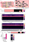
Mucin-related glycosyl hydrolase profiles in the Verrucomicrobia and Lentisphaerae phlyum. (A) Representative intestinal mucin glycans structures and corresponding microbial GHs. (B) Heat map of the percentage of Akkermansia glycaniphila or Akkermansia muciniphila genomes that have at least one gene copy of mucin-associated GH mucin-associated GH 33, 16, 29, 95, 20, 2, 35, 42, 98, 101, 129, 89, 85, and 84. (C) Heat maps depicting the gene copy number of mucin-associated GHs in the strains of A. glycaniphila and A. muciniphila. (D) Growth analysis of A. muciniphila ATCC BAA-835 in a chemically defined media ZMB1 lacking glucose (media control), with glucose (positive control), or lacking glucose and supplemented with 1 mg/mL porcine intestinal MUC2. Growth was measured by examining the optical density at 600 nm (OD600nm) after overnight incubation. (E) Heat maps showing the percentage of genomes that have at least one gene copy of each mucin-associated GH and depicting the gene copy number of mucin-associated GHs in the one strain of Victivallales bacterium.
Figure 3
Figure 3
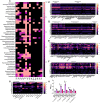
Mucin-related glycosyl hydrolase profiles in the Actinobacteria phlyum. (A) Heat map of the Actinobacteria genomes that have at least one gene copy of a mucin-associated GH 33, 16, 29, 95, 20, 2, 35, 42, 98, 101, 129, 89, 85, and 84. (B) Heat map showing the gene copy number of mucin-associated GHs in the strains of Actinomadura and Actinomyces, (C) Bifidobacteria, specifically B. bifidum and B. breve, (D) B. longum (Bl), B. longum subsp. infantis (Bli), B. longum subsp. longum (Bll), B. longum subsp. suillum (Bls), and B. scardovii, (E) Streptacidiphilus and Streptomyces species, and (F) Streptomyces species. (G) Growth analysis of Bifidobacterium dentium ATCC 27678, B. longum subsp. infantis ATCC 15697, B. bifidum ATCC 29521, B. longum ATCC 55813, and B. angulatum ATCC 27535 in a chemically defined media ZMB1 lacking glucose (media control), with glucose (positive control), or lacking glucose and supplemented with 1 mg/mL porcine intestinal MUC2. Growth was measured by examining the optical density at 600 nm (OD600nm) after overnight incubation.
Figure 4
Figure 4
Mucin-related glycosyl hydrolase profiles in the Bacteroidetes phlyum. (A) Heat map of the genera within the Bacteroidetes phlyum that have at least one gene copy of each mucin-associated GH 33, 16, 29, 95, 20, 2, 35, 42, 98, 101, 129, 89, 85, and 84. Heat map showing the gene copy number of mucin-associated GHs in the strains of (B) Alistipes, Alloprevotella, and Fermenitomonas, (C) Bacteroides, specifically B. caccae, B. dorei, B. intestinalis, B. fragilis and B. ovatus, (D) Bacteroides, specifically Bacteroides spp., B. thetaiotaomicron, B. uniformis, and B. xylanisolvens, (E) Prevotella copri, P. jejuni, and P melaninogenica, (F) Parabacteroides and P. distasonis, and (G) Phocaeicola coprophilus, P. dorei, and P. vulgatus. (H) Growth analysis of Bacteroides vulgatus ATCC 8482, B. thetaiotaomicron ATCC 29148, B. fragilis MGH 10513, Prevotella merdae MGH 10511, and Prevotella copri DSMZ 18205 in a chemically defined media ZMB1 lacking glucose (media control), with glucose (positive control), or lacking glucose and supplemented with 1 mg/mL porcine intestinal MUC2. Growth was measured by examining the optical density at 600 nm (OD600nm) after overnight incubation.
Figure 5
Figure 5
Mucin-related glycosyl hydrolase profiles in the Proteobacteria phlyum. (A) Heat map of the Proteobacteria genomes that have at least one gene copy of each mucin-associated GH 33, 16, 29, 95, 20, 2, 35, 42, 98, 101, 129, 89, 85, and 84. Heat map showing the gene copy number of mucin-associated GHs in the strains of (B) Klebsiella aerogenes, (C) Klebsiella spp., (D) Mixta calida, M. intestinalis, and Serratia fonticola, (E) Enterobacter cloacae, (F) Enterobacter spp. and E. asburiae. (G) Growth analysis of E. coli Nissle 1917 in a chemically defined media ZMB1 lacking glucose (media control), with glucose (positive control), or lacking glucose and supplemented with 1 mg/mL porcine intestinal MUC2. Growth was measured by examining the optical density at 600 nm (OD600nm) after overnight incubation.
Figure 6
Figure 6
Mucin-related glycosyl hydrolase profiles in the Firmicutes phlyum. (AC) Heat map of the Firmicutes genomes that have at least one gene copy of mucin-associated GH 33, 16, 29, 95, 20, 2, 35, 42, 98, 101, 129, 89, 85, and 84. Heat map showing the gene copy number of mucin-associated GHs in the strains of (D) Abiotrophia defective, Blautia coccoides, B. hansenii, B. obeum, B. producta, Blautia spp., Enterococcus casseliflavus, E. durans, E. gallinarum, and Enterococcus spp., (E) Paenibacillus, specifically Paenibacillus spp., P barcinonensis and P. lautus, (D) Ruminococcus, including Ruminococcus spp. R. gnavus and R. torques, (F) Streptococcus, including S. australis (Sa), S. intermedius (Si), S. mitis (Sm), Streptococcus spp. and Viridibacillus spp. (G) Clostridium, including C. butyricum (Cb), C. sporogenes (Cs), and Clostridium spp. (H,I) Growth analysis of Clostridium butyricum CB, Clostridium symbiosum ATCC 14940, Clostridium inoculum ATCC 14501, Clostridium clostridiforme ATCC 25532, and Clostridium sporogenes DSMZ 795 (H), as well as Lactobacillus gasseri ATCC 33323, L. johnsonii ATCC 33200, L. brevis ATCC 27305, L. acidophilus ATCC 4796 (I) in a chemically defined media ZMB1 lacking glucose (media control), with glucose (positive control), or lacking glucose and supplemented with 1 mg/mL porcine intestinal MUC2. Growth was measured by examining the optical density at 600 nm (OD600nm) after overnight incubation.

Similar articles

Cited by

References

    1. Corfield AP, Wagner SA, Clamp JR, Kriaris MS, Hoskins LC. Mucin degradation in the human colon: Production of sialidase, sialate O-acetylesterase, N-acetylneuraminate lyase, arylesterase, and glycosulfatase activities by strains of fecal bacteria. Infect. Immun. 1992;60:3971–3978. doi: 10.1128/iai.60.10.3971-3978.1992. - DOI - PMC - PubMed
    1. Bell A, Juge N. Mucosal glycan degradation of the host by the gut microbiota. Glycobiology. 2021;31:691–696. doi: 10.1093/glycob/cwaa097. - DOI - PMC - PubMed
    1. Tailford LE, Crost EH, Kavanaugh D, Juge N. Mucin glycan foraging in the human gut microbiome. Front. Genet. 2015;6:81. doi: 10.3389/fgene.2015.00081. - DOI - PMC - PubMed
    1. McGuckin MA, Linden SK, Sutton P, Florin TH. Mucin dynamics and enteric pathogens. Nat. Rev. Microbiol. 2011;9:265–278. doi: 10.1038/nrmicro2538. - DOI - PubMed
    1. McDole JR, et al. Goblet cells deliver luminal antigen to CD103+ dendritic cells in the small intestine. Nature. 2012;483:345–349. doi: 10.1038/nature10863. - DOI - PMC - PubMed

LinkOut - more resources