. 2022 May 19;13(1):2784.
doi: 10.1038/s41467-022-30448-8.

Templated folding of the RTX domain of the bacterial toxin adenylate cyclase revealed by single molecule force spectroscopy

Affiliations

Templated folding of the RTX domain of the bacterial toxin adenylate cyclase revealed by single molecule force spectroscopy

Han Wang et al. Nat Commun. .

Abstract

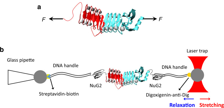
The RTX (repeats-in-toxin) domain of the bacterial toxin adenylate cyclase (CyaA) contains five RTX blocks (RTX-i to RTX-v) and its folding is essential for CyaA's functions. It was shown that the C-terminal capping structure of RTX-v is critical for the whole RTX to fold. However, it is unknown how the folding signal transmits within the RTX domain. Here we use optical tweezers to investigate the interplay between the folding of RTX-iv and RTX-v. Our results show that RTX-iv alone is disordered, but folds into a Ca2+-loaded-β-roll structure in the presence of a folded RTX-v. Folding trajectories of RTX-iv-v reveal that the folding of RTX-iv is strictly conditional upon the folding of RTX-v, suggesting that the folding of RTX-iv is templated by RTX-v. This templating effect allows RTX-iv to fold rapidly, and provides significant mutual stabilization. Our study reveals a possible mechanism for transmitting the folding signal within the RTX domain.


Conflict of interest statement

The authors declare no competing interests.

Figures

Fig. 1
Fig. 1. Probing the unfolding and folding of RTX-iv using OT.
a The 3D structure of RTX-iv-v (PDB: 6SUS). RTX-iv is colored in red and RTX-v is colored in cyan. Ca2+ ions are shown as gray spheres. RTX-iv and RTX-v fold into a seemingly continuous β-roll structure. b Schematics of the OT experiment on NuG2-RTX-iv-v-NuG2. The protein chimera NuG2-RTX-iv-v-NuG2 is coupled with two DNA handles via thiol-maleimide chemistry. The DNA–protein complex is then attached to two polystyrene beads, which are functionalized with streptavidin and anti-digoxigenin, via specific ligand-receptor interactions. One bead is held by a glass micropipette and the other is trapped in the laser trap. By moving the position of laser trap, the target protein can be mechanically stretched or relaxed, and the force–distance relationship of the protein-DNA chimera can be measured.
Fig. 2
Fig. 2. RTX-iv alone is intrinsically disordered.
a Far UV CD spectrum of RTX-iv in the absence of Ca2+ and the presence of 10 mM Ca2+. b Representative F–D curves of NuG2-RTX-iv-NuG2 in Tris-HCl (left) and Tris-HCl+10 mM Ca2+ (right) at a pulling speed of 50 nm/s. Only the unfolding and refolding events of NuG2 domains were observed, suggesting that RTX-iv alone behaved as a disordered polypeptide.
Fig. 3
Fig. 3. Holo-RTX-iv-v is mechanically stable and can fold rapidly.
a Representative F–D curves of the full-length NuG2-RTX-iv-v-NuG2. The unfolding and refolding events of NuG2 domains are colored in black and indicated by the squares, and the unfolding–folding of RTX-iv and RTX-v are colored in red and cyan, respectively. b Representative stretching and relaxation F–D curves showing the unfolding–folding of RTX-iv and RTX-v. The unfolding and folding events of RTX-iv are colored in red and indicated by circles, and the unfolding–folding events of RTX-v are colored in cyan and indicated by squares. For clarity, the two stretching–relaxation cycles are offset relative to each other. c F–D curves show that unfolding and folding of RTX-iv occur via a four-state pathway involving two intermediate states. The stretching–relaxation curves shown herein were limited to below 11–12 pN so that only RTX-iv unfolds and refolds, while RTX-v remained folded throughout. Insets show the zoomed view of the F–D curves showing the four-state unfolding and folding. Gray dashed lines in (b, c) are pseudo-worm-like chain (WLC) fits to the F–D curves. d Force–extension relationships of the (un)folding of RTX-iv-v. WLC fits (dashed lines) to the experimental data measured a ΔLc-RTX-iv of 39.9 ± 0.8 nm (n = 37) and ΔLc-RTX-v of 46.2 ± 0.9 nm (n = 42), respectively. WLC fits to the unfolding events involving intermediate states revealed ΔLc1 of 6.5 ± 0.9 nm (n = 87, from the native state to the first intermediate state, N-I1), ΔLc2 of 11.3 ± 0.7 nm (n = 91, from the first intermediate state to the second intermediate state, I1–I2), ΔLc3 of 23.5 ± 0.5 nm (n = 115, from the second intermediate state to the unfolded state, I2-U), respectively. The error bars indicate standard deviations. e Histograms of unfolding and refolding forces of RTX-iv at a pulling speed of 50 nm/s. The number of unfolding and refolding events is shown in Supplementary Table 1.
Fig. 4
Fig. 4. Mechanical unfolding/folding of RTX-iv-T1411C-v.
a Schematics of stretching RTX-iv-T1411C-v from the C-terminus and T1411C (indicated by the green asterisk). b Representative F–D curves of RTX-iv-T1411C-v, in which the molecule was stretched to unfold–fold RTX-iv-T1411C only. The unfolding and folding patterns are the same as those of wt RTX-iv. c Force–extension relationships of the unfolding of RTX-iv-T1411C. The WLC fits yielded a ΔLc1 (n = 57, N-I1) of 6.1 ± 0.4 nm, ΔLc2 (n = 52, I1–I2) of 11.0 ± 0.5 nm, ΔLc3 (n = 64, I2-U) of 23.1 ± 0.6 nm and ΔLc (n = 30, RTX-iv-T1411C) of 39.7 ± 0.8 nm. The error bars indicate standard deviations.
Fig. 5
Fig. 5. The folding of RTX-iv is conditional upon the folding of the whole RTX-v.
a Schematics of the structure of RTX-v50. RTX-v50 is constructed by inserting an intrinsically disordered elastin-like sequence (VPGAG)10 between residue Leu1572 and Ser1573 in wt RTX-v. b A representative stretching–relaxation F–D curve of RTX-iv-v50. The unfolding event of RTX-iv is colored in red, and the unfolding and partial refolding events of RTX-v50 are colored in cyan. c, d Consecutive F–D curves showing the unfolding and refolding of RTX-iv-v50. The unfolding event of RTX-iv is colored in red, and unfolding and refolding events of RTX-v50 are colored in cyan. c No waiting time (Δt) was allowed between consecutive stretching–relaxation cycles, while in (d) Δt was 60 s. In the relaxation curves of RTX-iv-v50, only the refolding of C-FI at ∼4 pN was observed, while no trajectory in which both RTX-iv and the N-terminal part of RTX-v refolded was observed. e The relationship of folding probability versus Δt at zero force of the N-terminal part of RTX-v50. Fitting the data to the first-order kinetics led to a folding rate constant of (2.1 ± 0.4) × 10−2 s−1 of the N-terminal part of RTX-v50 at zero force. The number of events at Δt = 0, 5, 10, 30, 60 s are 172, 47, 35, 57, and 24, respectively. The error bars indicate standard deviations.
Fig. 6
Fig. 6. Mechanical unfolding/folding of RTX-iv-T1483C-v.
a Schematics of stretching RTX-iv-T1483C-v from its C-terminus and the residue 1483 (indicated by the green asterisk). b Representative force–distance curves showing the unfolding/folding of RTX-iv-T1483C-v at a pulling speed of 50 nm/s. The unfolding/folding of RTX-iv-T1483C is colored in red, and the unfolding/folding of RTX-v is colored in cyan. Both unfolding and folding initiate from RTX-v. c The WLC fitting of the unfolding of force–extension relationships of the unfolding of RTX-iv-T1483C (red) and RTX-v (cyan), respectively. The WLC fits yielded a ΔLc of 16.1 ± 0.3 nm (n = 105) for RTX-iv-T1483C, and ΔLc of 45.2 ± 0.7 nm (n = 105) for RTX-v, respectively. The error bars indicate standard deviations.

Similar articles

References

    1. Fernández-Puentes C, Carrasco L. Viral infection permeabilizes mammalian cells to protein toxins. Cell. 1980;20:769–775. doi: 10.1016/0092-8674(80)90323-2. - DOI - PubMed
    1. Montecucco C, Papini E, Schiavo G. Bacterial protein toxins penetrate cells via a four-step mechanism. FEBS Lett. 1994;346:92–98. doi: 10.1016/0014-5793(94)00449-8. - DOI - PubMed
    1. Falnes PO, Sandvig K. Penetration of protein toxins into cells. Curr. Opin. Cell. Biol. 2000;12:407–413. doi: 10.1016/S0955-0674(00)00109-5. - DOI - PubMed
    1. Parker MW, Feil SC. Pore-forming protein toxins: from structure to function. Prog. Biophys. Mol. Biol. 2005;88:91–142. doi: 10.1016/j.pbiomolbio.2004.01.009. - DOI - PubMed
    1. O'Brien DP, et al. Post-translational acylation controls the folding and functions of the CyaA RTX toxin. FASEB J. 2019;33:10065–10076. doi: 10.1096/fj.201802442RR. - DOI - PubMed

Publication types

MeSH terms

Grants and funding