Review
. 2022 Dec;23(12):797-816.
doi: 10.1038/s41580-022-00490-x. Epub 2022 May 19.

Phosphoinositides as membrane organizers

Affiliations
Review

Phosphoinositides as membrane organizers

York Posor et al. Nat Rev Mol Cell Biol. 2022 Dec.

Abstract

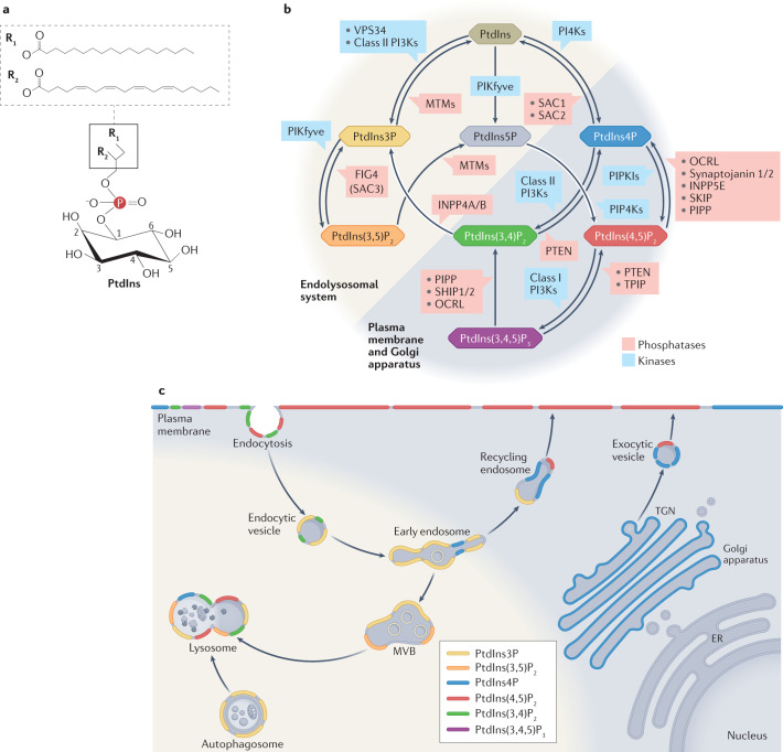
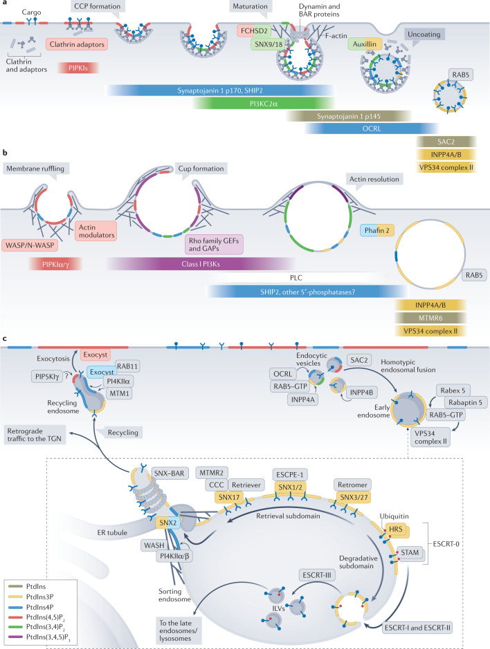
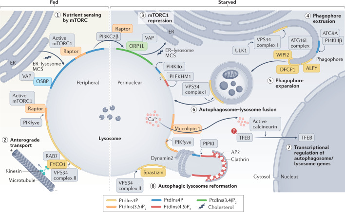
Phosphoinositides are signalling lipids derived from phosphatidylinositol, a ubiquitous phospholipid in the cytoplasmic leaflet of eukaryotic membranes. Initially discovered for their roles in cell signalling, phosphoinositides are now widely recognized as key integrators of membrane dynamics that broadly impact on all aspects of cell physiology and on disease. The past decade has witnessed a vast expansion of our knowledge of phosphoinositide biology. On the endocytic and exocytic routes, phosphoinositides direct the inward and outward flow of membrane as vesicular traffic is coupled to the conversion of phosphoinositides. Moreover, recent findings on the roles of phosphoinositides in autophagy and the endolysosomal system challenge our view of lysosome biology. The non-vesicular exchange of lipids, ions and metabolites at membrane contact sites in between organelles has also been found to depend on phosphoinositides. Here we review our current understanding of how phosphoinositides shape and direct membrane dynamics to impact on cell physiology, and provide an overview of emerging concepts in phosphoinositide regulation.


Conflict of interest statement

The authors declare no competing interests.

Figures

Fig. 1
Fig. 1. Metabolism and subcellular enrichment of phosphoinositides.
a | Chemical structure of phosphatidylinositol (PtdIns), a glycerophospholipid with myo-inositol as a head group. Note that only the 2′-hydroxy group of the inositol ring is in the axial position (that is, perpendicular to the plane of the inositol ring), while all others are equatorial. PtdIns is depicted in the 1-stearoyl (R1), 2-arachidonyl (R2) configuration, the most frequent acyl chain composition found in phosphoinositides. b | Phosphoinositide metabolism. Addition and removal of phosphate groups at the 3′-hydroxy, 4′-hydroxy and 5′-hydroxy groups of PtdIns by phosphoinositide kinases (blue) and phosphatases (red) creates seven distinct phosphoinositide species. Enzymatic interconversion of phosphoinositides creates a network in which formation of one species depends on the availability of another, meaning that the cellular levels of certain phosphoinositides are interdependent. Phosphoinositides enriched in the endolysosomal system (shaded yellow) versus the secretory pathway and the plasma membrane (shaded blue) are indicated. c | Subcellular enrichment of phosphoinositides. The spatiotemporally controlled activity of phosphoinositide kinases and phosphatases creates a distinctive enrichment of phosphoinositides across the cellular compartments. The plasma membrane is highly enriched in PtdIns 4,5-bisphosphate (PtdIns(4,5)P2). The Golgi apparatus and the trans-Golgi network (TGN) are characterized by PtdIns 4-phosphate (PtdIns4P), which is also present at the plasma membrane. By contrast, the endosomal compartments are dominated by 3′-phosphoinositides, with PtdIns 3-phosphate (PtdIns3P) being the signature lipid of early endosomes. A fraction of PtdIns3P is converted to PtdIns 3,5-bisphosphate (PtdIns(3,5)P2) on late endosomes. Lysosomes are unique in having a diverse complement of phosphoinositides in their membranes, likely depending on their functional and metabolic state. On multiple endocytic routes, PtdIns 3,4-bisphosphate (PtdIns(3,4)P2) is formed on nascent endocytic carriers before conversion to PtdIns3P in endocytic vesicles. ER, endoplasmic reticulum; MTM, myotubularin; MVB, multivesicular body; PI3K; phosphatidylinositol 3-kinase; PI4K, phosphatidylinositol 4-kinase; PIPKI, phosphatidylinositol 4-phosphate 5-kinase; PtdIns(3,4,5)P2, phosphatidylinositol 3,4,5-trisphosphate; PtdIns5P, phosphatidylinositol 5-phosphate.
Fig. 2
Fig. 2. Phosphoinositide conversions during endocytosis and endosomal sorting.
a | In clathrin-mediated endocytosis, nucleation and growth of endocytic clathrin-coated pits (CCPs) are promoted by phosphatidylinositol 4,5-bisphosphate (PtdIns(4,5)P2). Many adaptors, which connect the clathrin lattice with the membrane and concentrate cargo proteins in the nascent pit, bind to PtdIns(4,5)P2. Maturing CCPs acquire a number of phosphoinositide-metabolizing enzymes, including PI3KC2α and the 5-phosphatases SHIP2 and synaptojanin 1 (p170). The resulting formation of phosphatidylinositol 3,4-bisphosphate (PtdIns(3,4)P2) through its effectors sorting nexin 9 (SNX9), SNX18 and FCHSD2 promotes constriction of the neck of invaginated CCPs. During uncoating of newly formed vesicles, a burst of recruitment of the 5-phosphatases synaptojanin 1 (p145) and OCRL depletes PtdIns(4,5)P2. The 4-phosphatases INPP4A, INPP4B and SAC2 complete conversion of membrane identity towards the endosomal signature lipid phosphatidylinositol 3-phosphate (PtdIns3P). b | Macropinocytosis, the internalization of large amounts of extracellular fluid, is largely driven by membrane remodelling through branched F-actin formation. Initial membrane ruffling requires the formation of PtdIns(4,5)P2 by PIPKIα/γ to promote actin polymerization. Receptor (that is, insulin or growth factor receptors) activation triggers the formation of phosphatidylinositol 3,4,5-trisphosphate (PtdIns(3,4,5)P3) by class I phosphatidylinositol 3-kinases (PI3Ks) to regulate Rho family guanine-nucleotide exchange factors (GEFs) and GTPase-activating proteins (GAPs) during expansion of the macropinocytic cup. Remodelling and resolution of the subcortical actin cytoskeleton requires depletion of PtdIns(4,5)P2 by phospholipase C (PLC) and 5-phosphatases starting from the base of the cup. These phosphoinositide conversions enable recruitment of the dual PtdIns3P- and phosphatidylinositol 4-phosphate (PtdIns4P)-binding protein Phafin 2, a protein also required for actin remodelling. c | The endosomal sorting of cargo depends on PtdIns3P. Endosomal PtdIns3P is largely synthesized by VPS34 complex II, which is activated by RAB5–GTP. Coincidence of cargo and PtdIns3P underlies the sorting of cargo into degradative and retrieval subdomains. This is mediated by proteins that can simultaneously bind to PtdIns3P and to specific sorting signals in cargo proteins. Endosomal sorting complex required for transport (ESCRT) 0 binds to ubiquitylated proteins and through self-association forms degradative subdomains. Retrieval signals are recognized by various members of the SNX family that operate in conjunction with different retrieval complexes. Exit from endosomes and recycling to the plasma membrane require conversion of PtdIns3P to PtdIns4P (and possibly PtdIns(4,5)P2). Proteins that interact with phosphoinositides are highlighted with coloured boxes, corresponding to the phosphoinositide species they bind to. The coloured bars highlight the phosphoinositide kinases and phosphatases, with colours corresponding to the phosphoinositide species they generate. BAR, Bin–Amphiphysin–Rvs; ER, endoplasmic reticulum; ILV, intraluminal vesicle; MTM1, myotubularin 1; MTMR, myotubularin-related protein; PIPKI, phosphatidylinositol 4-phosphate 5-kinase; PtdIns, phosphatidylinositol; TGN, trans-Golgi network; WASH, Wiskott-Aldrich syndrome protein and SCAR homologue.
Fig. 3
Fig. 3. Phosphoinositide control of lysosome function.
Lysosomes harbour a diverse complement of phosphoinositides that regulate their functions. In fed cells (left), lysosomal phosphatidylinositol 3-phosphate (PtdIns3P) and phosphatidylinositol 3,5-bisphosphate (PtdIns(3,5)P2) synthesized by VPS34 and PIKfyve, respectively, facilitate nutrient signalling via mechanistic target of rapamycin complex 1 (mTORC1), possibly by associating with its regulatory associated protein of mTOR (Raptor) subunit (number 1). mTORC1 activity is facilitated by oxysterol-binding protein (OSBP)-mediated cholesterol transfer from the endoplasmic reticulum (ER) at membrane contact sites (MCSs). PtdIns3P also promotes the anterograde transport of lysosomes via kinesin recruitment by FYVE and coiled-coil domain-containing protein 1 (FYCO1) and its binding partner RAB7 (number 2). Under starvation (right), mTORC1 activity is repressed by local phosphatidylinositol 3,4-bisphosphate (PtdIns(3,4)P2) synthesis mediated by class II phosphatidylinositol 3-kinase-β (PI3KC2β). PtdIns(3,4)P2 facilitates reverse cholesterol transfer from lysosomes to the ER via OSBP-related protein 1 long isoform (ORP1L) at MCSs (number 3). Concomitantly, starvation induces autophagy. Delivery of vesicles containing PI4KIIIβ and ATG9A may aid phagophore membrane extrusion (number 4). Formation of PtdIns3P by VPS34 complex I is required to drive expansion of the phagophore membrane from the ER by recruiting multiple effector proteins, leading to LC3 lipidation by the ATG16L complex (ATG12–ATG5–ATG16L) and autophagosome formation (number 5). Membrane fusion between PtdIns3P-containing autophagosomes and lysosomes involves complex pathways of PtdIns3P–PtdIns(3,5)P2 and phosphatidylinositol 4-phosphate (PtdIns4P)–phosphatidylinositol 4,5-bisphosphate (PtdIns(4,5)P2) synthesis and turnover that involve phosphatidylinositol 4-kinase IIα (PI4KIIα) and the RAB7 effector protein PLEKHM1 (number 6). Starvation-triggered Ca2+ efflux via the PtdIns(3,5)P2-activated Ca2+-channel mucolipin 1 activates calcineurin and, thereby, induces transcription of autophagy/lysosomal genes via nuclear translocation of transcription factor EB (TFEB) (number 7). Prolonged starvation triggers autophagic lysosome reformation (ALR), a process that involves multiple lysosomal phosphoinositides. Synthesis of PtdIns(4,5)P2 on lysosomes by PtdIns4P 5-kinase (PIPKI) isoforms induces the formation of lysosomal tubules and the assembly of clathrin–AP2 coats that support the budding of protolysosomes from tubular intermediates via dynamin 2. Tubulation is further facilitated by VPS34 complex II-mediated synthesis of PtdIns3P, which serves to recruit the FYVE domain-containing protein spastizin, and by PtdIns(3,5)P2 synthesis via PIKfyve (number 8). The colour of proteins indicates the phosphoinositide species they bind to. ALFY, autophagy-linked FYVE protein; DFCP1, double FYVE domain-containing protein 1; ULK1, UNC-51-like autophagy-activating kinase 1; VAP, VAMP-associated protein; WIPI2, WD repeat domain phosphoinsitide-interacting protein 2.
Fig. 4
Fig. 4. Phosphoinositides at membrane contact sites.
Phosphoinositides regulate the formation of membrane contact sites (MCSs) (panel a), while MCSs mediate lipid transfer and control phosphoinositide metabolism (panel b). a | During MCS formation, phosphoinositides in the membrane of one organelle recruit a specific phosphoinositide-binding protein anchored in the membrane of another organelle. Examples of this organization include endoplasmic reticulum (ER)–plasma membrane (PM) contacts mediated by extended synaptotagmin 1 (E-Syt1) (number 1); ER–trans-Golgi network (TGN) MCSs formed by ceramide transfer protein (CERT) and VAMP-associated protein (VAP) (number 2); ER–late endosome MCSs formed by protrudin with PDZD8, VAP and RAB7 (number 3); ER–lysosome MCSs formed by oxysterol-binding protein (OSBP)-related protein 1 long isoform (ORP1L) and VAP; and ER–omegasome contacts containing WD repeat domain phosphoinositide-interacting protein 4 (WIPI4) and autophagy-related protein ATG2. b | Vectorial transfer of lipids at MCSs. Lipid transfer can follow the concentration gradient or can be powered by the countertransport of another lipid. Examples of this lipid transfer include countertransport of cholesterol and phosphatidylinositol 4-phosphate (PtdIns4P) by OSBP at MCSs between the ER and the TGN (number 1); phosphatidylserine (PtdSer) and PtdIns4P by ORP10 at ER–endosome contacts (number 2); and PtdSer and PtdIns4P/phosphatidylinositol 4,5-bisphosphate (PtdIns(4,5)P2) by OSBP-related protein 5 (ORP5)/ORP8 at ER–plasma membrane MCSs (number 3). In addition, during infection with hepatitis C virus (HCV), Nir2 catalyses transport of phosphatidylinositol (PtdIns) from the ER to the plasma membrane, the Golgi complex, or HCV replicative organelles (HCV-RO) in exchange for phosphatidic acid (PtdOH) (number 4). In pancreatic β-cells, TMEM24 replenishes plasma membrane PtdIns lost during glucose-stimulated phospholipase C (PLC) signalling under conditions of low Ca2+ levels. PtdIns shuttled via Nir2 or TMEM24 is phosphorylated by phosphatidylinositol 4-kinase (PI4K) and PtdIns4P 5-kinase (PI4P5K) at the plasma membrane. FYVE, phosphatidylinositol 3-phosphate-binding domain named after FAB1, YOTB, VAC1 and EEA1; ORD, OSBP-related domain; PITP, phosphatidylinositol transfer protein domain; PH, pleckstrin homology domain; PtdIns3P, phosphatidylinositol 3-phosphate; PtdIns(3,4)P2, phosphatidylinositol 3,4-bisphosphate; PtdIns(3,4,5)P3, phosphatidylinositol 3,4,5-trisphosphate; SMP, synaptotagmin-like mitochondrial lipid-binding domain; StART, StAR-related lipid transfer domain.

Similar articles

Cited by

References

    1. Behnia R, Munro S. Organelle identity and the signposts for membrane traffic. Nature. 2005;438:597–604. doi: 10.1038/nature04397. - DOI - PubMed
    1. Prinz WA, Toulmay A, Balla T. The functional universe of membrane contact sites. Nat. Rev. Mol. Cell Biol. 2020;21:7–24. doi: 10.1038/s41580-019-0180-9. - DOI - PMC - PubMed
    1. Wu H, Carvalho P, Voeltz GK. Here, there, and everywhere: the importance of ER membrane contact sites. Science. 2018 doi: 10.1126/science.aan5835. - DOI - PMC - PubMed
    1. Balla T. Phosphoinositides: tiny lipids with giant impact on cell regulation. Physiol. Rev. 2013;93:1019–1137. doi: 10.1152/physrev.00028.2012. - DOI - PMC - PubMed
    1. Bilanges B, Posor Y, Vanhaesebroeck B. PI3K isoforms in cell signalling and vesicle trafficking. Nat. Rev. Mol. Cell Biol. 2019 doi: 10.1038/s41580-019-0129-z. - DOI - PubMed

Publication types

MeSH terms

Substances