Review
. 2022 May 19;79(6):304.
doi: 10.1007/s00018-022-04314-1.

Regulation of choroid plexus development and its functions

Affiliations
Review

Regulation of choroid plexus development and its functions

Petra Kompaníková et al. Cell Mol Life Sci. .

Abstract

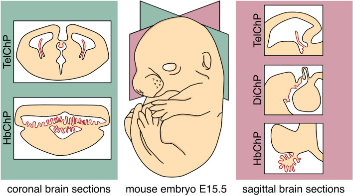
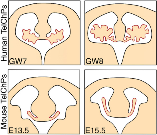
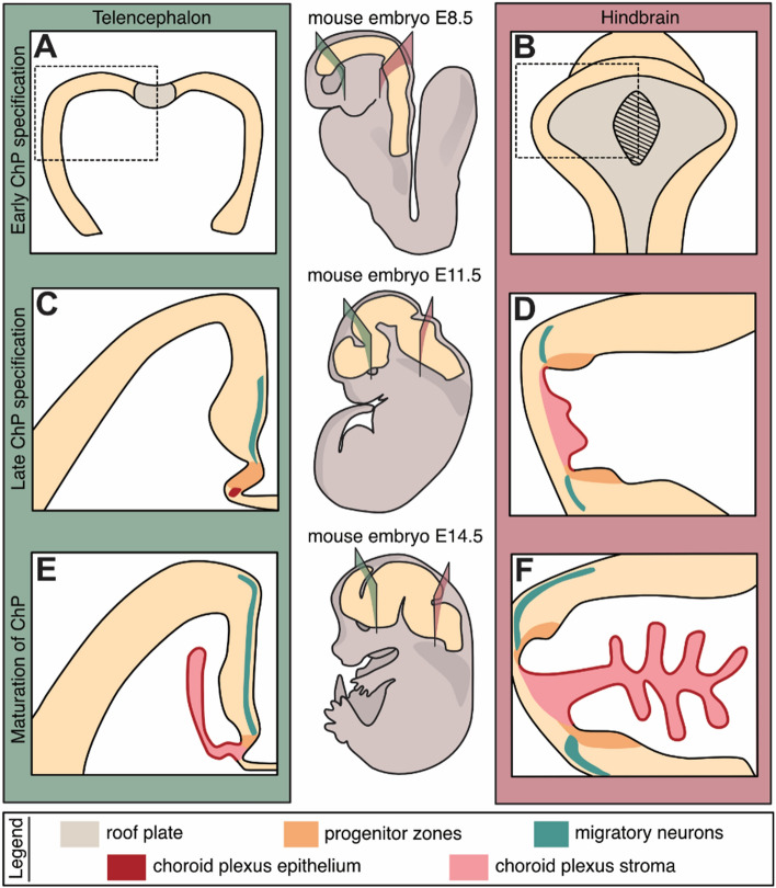
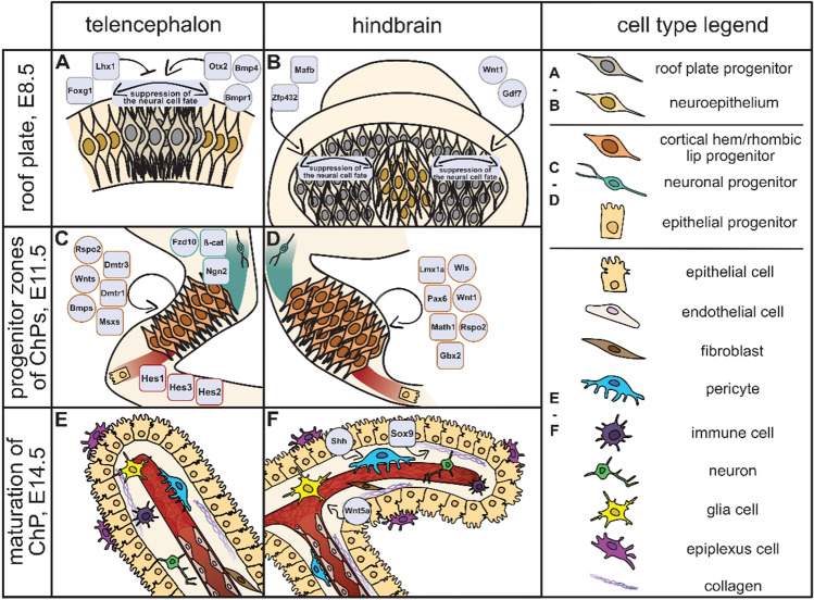
The choroid plexus (ChP) is an extensively vascularized tissue that protrudes into the brain ventricular system of all vertebrates. This highly specialized structure, consisting of the polarized epithelial sheet and underlying stroma, serves a spectrum of functions within the central nervous system (CNS), most notably the production of cerebrospinal fluid (CSF). The epithelial cells of the ChP have the competence to tightly modulate the biomolecule composition of CSF, which acts as a milieu functionally connecting ChP with other brain structures. This review aims to eloquently summarize the current knowledge about the development of ChP. We describe the mechanisms that control its early specification from roof plate followed by the formation of proliferative regions-cortical hem and rhombic lips-feeding later development of ChP. Next, we summarized the current knowledge on the maturation of ChP and mechanisms that control its morphological and cellular diversity. Furthermore, we attempted to review the currently available battery of molecular markers and mouse strains available for the research of ChP, and identified some technological shortcomings that must be overcome to accelerate the ChP research field. Overall, the central principle of this review is to highlight ChP as an intriguing and surprisingly poorly known structure that is vital for the development and function of the whole CNS. We believe that our summary will increase the interest in further studies of ChP that aim to describe the molecular and cellular principles guiding the development and function of this tissue.

Keywords: Cerebrospinal fluid (CSF); ChP epithelial cells; Choroid plexus (ChP); Cortical hem; Morphogenesis; Rhombic lips.


Conflict of interest statement

The authors declare no conflict of interest.

Figures

Fig. 1
Fig. 1
The localization of the ChP secretory system within the developing mouse brain. The set of three distinct ChP types (highlighted in red colour) occupies every ventricle formed within the main brain embryonic regions. This includes lateral ventricles marked by the presence of telencephalic TelChPs, 3rd ventricle characterized by the small DiChP and 4th ventricle fills with the robust HbChP. A growing body of evidence indicated differences between the individual types of ChP, not only in their diverse morphological appearance, which can be appreciated on the coronal (panel in dark green) as well as sagittal sections (panel in dark pink) of the murine brain, but also in the expression profiles of the ChP epithelia. This spatial regionalization translates into differences in the composition of the cerebrospinal fluid (CSF) which ChPs produces into the lumen of the ventricular system. ChP choroid plexus, TelChP telencephalic choroid plexus, DiChP diencephalic choroid plexus, HbChP hindbrain choroid plexus, CSF cerebrospinal fluid (CSF), E embryonic day
Fig. 2
Fig. 2
The distinct morphological shapes of TelChPs in mouse and human. The TelChPs outgrows gradually out of the medial telencephalic wall into the lateral ventricles. TelChPs fill up to 63% of the total cavity area in humans [16] while the murine TelChPs occupied only 27% of the mapped area [17]. These differences are due to a more complicated morphology of human TelChPs. TelChP telencephalic choroid plexus, GW gestation week, E embryonic day
Fig. 3
Fig. 3
The ontogenesis of ChPs. A schematic simplification of ChP development and formation is shown on the coronal brain sections of mouse telencephalon (panels in green, left) and hindbrain (panels in dark pink, right) at E8.5 (A, B), E11.5 (C, D) and E14.5 (E, F). The early specification of the TelChP as well as HbChP epithelial cells (ChPEC) (highlighted in red within panels CF), takes place in the embryonic roof plate (in grey; except the hatched part in B) (A, B). Further development (panels C,D as close up of neural tube regions indicated by dashed boxes in A, B) of the ChPs is connected with the formation of progenitor domains (orange). They are called cortical hem (CH) in the telencephalon (C) and rhombic lips (RL) in the hindbrain (D). These regions, besides ChPEC, also give rise to the migratory neurons (green). They are represented by Cajal–Retzius cells (from CH) migrating into the developing cortex of the telencephalon (C, E) as well as hindbrain migratory neurons targeting the cerebellum (from upper RL) (D, F). The last step in the ChP ontogenesis is its maturation when ChP gets its typical shape (E, F). Maturation involves further differentiation of ChPEC and the formation of ChP stroma (pink) achieved by the migration of mesoderm-derived stromal cells
Fig. 4
Fig. 4
Cell types, transcription factors, and signalling pathways involved in the ontogenesis of ChPs. A summary of cell types and key molecular regulators involved in the development of ChP. Schematics directly connects to Fig. 3 that describes the roof plate (A, B; E8.5), progenitor zone (C, D; E11.5), and maturation (E, F; E14.5) stages of ChP development. The development of TelChP (A, C, E; left) and HbChP (B, D, F; right) is outlined separately. For the legend of individual cell types, see the panel on the right. Transcription factors are shown in rounded squares; components of morphogenetic signalling pathways are in circles. For further details and references, see the text in the chapter “Development of the ChP”

Similar articles

Cited by

References

    1. Brocklehurst G. The significance of the evolution of the cerebrospinal fluid system. Ann R Coll Surg Engl. 1979;61:349–356. - PMC - PubMed
    1. Davson H, Segal MB. Physiology of the CSF and blood-brain barriers. Boca Raton: CRC Press; 1996.
    1. Dohrmann GJ, Bucy PC. Human choroid plexus: a light and electron microscopic study. J Neltrosurg. 1970;33:506/516. doi: 10.3171/jns.1970.33.5.0506. - DOI - PubMed
    1. Dandy WE. Extirpation of the choroid plexus of the lateral ventricles in communicating hydrocephalus. Ann Surg. 1918;68:569–579. doi: 10.1097/00000658-191812000-00001. - DOI - PMC - PubMed
    1. Somerford AE. A case of papilloma of the choroid plexus. Arch Dis Child. 1933;8:53–56. doi: 10.1136/adc.8.43.53. - DOI - PMC - PubMed

LinkOut - more resources