. 2022 Oct;102(10):1132-1142.
doi: 10.1038/s41374-022-00795-7. Epub 2022 May 19.

Characterization and identification of measurable endpoints in a mouse model featuring age-related retinal pathologies: a platform to test therapies

Affiliations

Characterization and identification of measurable endpoints in a mouse model featuring age-related retinal pathologies: a platform to test therapies

Mayur Choudhary et al. Lab Invest. 2022 Oct.

Abstract

Apolipoprotein B100 (apoB100) is the structural protein of cholesterol carriers including low-density lipoproteins. It is a constituent of sub-retinal pigment epithelial (sub-RPE) deposits and pro-atherogenic plaques, hallmarks of early dry age-related macular degeneration (AMD), an ocular neurodegenerative blinding disease, and cardiovascular disease, respectively. Herein, we characterized the retinal pathology of transgenic mice expressing mouse apoB100 in order to catalog their functional and morphological ocular phenotypes as a function of age and establish measurable endpoints for their use as a mouse model to test potential therapies. ApoB100 mice were found to exhibit an age-related decline in retinal function, as measured by electroretinogram (ERG) recordings of their scotopic a-wave, scotopic b-wave; and c-wave amplitudes. ApoB100 mice also displayed a buildup of the cholesterol carrier, apolipoprotein E (apoE) within and below the supporting extracellular matrix, Bruch's membrane (BrM), along with BrM thickening, and accumulation of thin diffuse electron-dense sub-RPE deposits, the severity of which increased with age. Moreover, the combination of apoB100 and advanced age were found to be associated with RPE morphological changes and the presence of sub-retinal immune cells as visualized in RPE-choroid flatmounts. Finally, aged apoB100 mice showed higher levels of circulating and ocular pro-inflammatory cytokines, supporting a link between age and increased local and systemic inflammation. Collectively, the data support the use of aged apoB100 mice as a platform to evaluate potential therapies for retinal degeneration, specifically drugs intended to target removal of lipids from Bruch's membrane and/or alleviate ocular inflammation.


Conflict of interest statement

Disclosures/Conflict of Interest: JTH received grant funding and royalties from Bayer Pharmaceuticals, Inc, and grant funding and stock options as a member of the Scientific Advisory Board for Clover Pharmaceuticals, Inc, both for unrelated projects.

Conflict of interest statement: The authors have declared that no conflicts of interest exist.

Figures

Fig. 1
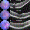
Fig. 1. In vivo imaging of apoB100 mouse eyes.
A Micron IV retinal imaging microscope was used to acquire fundus and OCT images. Representative images of apoB100 (a, b) 9-month old, Male, left eye (oculus sinister: OS), (c, d) 21-month old, Female, OS, and (e, f) 28-month old, Female, OS. OCT slice corresponds to the green arrow in the fundus images (INL: Inner nuclear layer, IPL: Inner plexiform layer, ONL: Outer nuclear layer, OPL: Outer plexiform layer, OS: Photoreceptor outer segments).
Fig. 2
Fig. 2. Retinal function of apoB100 mice with age.
Averaged ERG responses in 8–10 month old (black) and 17–21 month old (red) dark-adapted apoB100 mice (red) (n=5 per group). Plots of (a) photopic a-wave amplitudes, (b) photopic b-wave amplitudes, (c) photopic b-wave/a-wave ratio, (d) scotopic a–wave amplitudes and (e) scotopic b-wave amplitudes and (f) scotopic b-wave/a-wave ratio are shown as a function of flash intensity (For some points, the error bars are shorter than the height of the symbol). Data points are mean and SEM. * p<0.01. (a) Representative ERG traces from 8–10 month old (black) and 17–21 month old (red) dark-adapted apoB100 mice. (b) Averaged c-wave amplitudes in 8–10 month old (n=11) and 17–21 month old (n=5) apoB100 mice at a flash intensity of 0.2 cd.s/m2. Data points are mean ± SEM (8–12 month, n=11, 17–21 month, n=5, *p<0.05)
Fig. 3
Fig. 3. ApoE accumulates below the RPE and within BrM of aged apoB100 mice.
ApoE stained images of the outer retina of (a,b) 8–12 month old apoB100 mice and, (c, d) 17–21 month old apoB100 mice, displaying ApoE positive sub-RPE deposits and Bruch’s membrane thickening (Scale bar: 20 μm). Quantification of ApoE staining intensity in 8–12 month and 17–21 month old apoB100 mice (8–12 month, n=3, 17–21 month, n=4, *p<0.05).
Figure 4:
Figure 4:. Ultrastructural changes in apoB100 as a function of age.
(a, b) Low magnification TEM images from 8–12 month old apoB100 mice display regions with thin diffuse sub-RPE deposits and disrupted basal infoldings. (c, d) 17–21 month old mice exhibit continuous thin sub-RPE deposits and a higher degree of disrupted/absent basal infoldings (Scale bar = 2 μm). (e-f) High magnification images of 8–12 month old mice exhibit lipid droplets (asterisks) in BrM. (i-l) 17–21 month old mice display BrM thickening and sub-RPE deposits in addition to lipid droplets in the RPE as well as BrM (asterisk; Scale bar = 2 μm).
Fig. 5
Fig. 5. Distribution of sub-retinal immune cells and RPE degenerative changes in apoB100 mice increases in severity with age.
F4/80 immunoreactivity and Phallloidin stained images of RPE flatmounts from (a) 8–12 month old (Scale bar: 100 μm), (b) 17–21 month old, and, (c) 24–28 month old mice showing the distribution of immune cells in the outer retina and RPE morphology (ON: Optic nerve). (d) Quantification of F4/80 positive cells in apoB100 mice (8–12 month old, n=5, 17–21 month old, n=5 and 24–28 month old n=2, * p<0.05). Magnified images of RPE flatmounts (e) 8–12 month old (Scale bar: 20 μm), (f) 17–21 month old, and (g) 24–28 month old apoB100 mice showing RPE morphology and immune cell morphology. F4/80 stained images of retinal cross-sections from (h, i) 8–12 month old (Scale bar: 10 μm) and (j, K) 17–21 month old apoB100 mice displaying sub retinal immune cells (asterisks). (OPL: Outer plexiform layer, ONL: Outer nuclear layer, OS: photoreceptor outer segments, RPE: Retinal pigment epithelium).
Figure 6.
Figure 6.. Aged apoB100 mice exhibit a significant upregulation of proinflammatory cytokines in the systemic circulation.
Protein isolated from plasma and RPE-choroid tissue of 8–12 month and 17–21 month old mice were blotted on a Mouse Cytokine Array C3 (62 proteins). Representative images of significantly regulated cytokines are shown. (a, b) Plasma and (c, d) RPE-choroid. Quantification of staining intensity of significantly regulated cytokines. (n = 4 per group, Mean+SEM, *: p<0.05, Multiple t-tests).

Similar articles

Cited by

References

    1. Friedman DS, O’Colmain BJ, Munoz B, Tomany SC, McCarty C, de Jong PT et al. Prevalence of age-related macular degeneration in the United States. Arch Ophthalmol 122, 564–572 (2004). - PubMed
    1. Ambati J & Fowler BJ Mechanisms of age-related macular degeneration. Neuron 75, 26–39 (2012). - PMC - PubMed
    1. Li JQ, Welchowski T, Schmid M, Mauschitz MM, Holz FG & Finger RP Prevalence and incidence of age-related macular degeneration in Europe: a systematic review and meta-analysis. Br J Ophthalmol 104, 1077–1084 (2020). - PubMed
    1. Klein R, Peto T, Bird A & Vannewkirk MR The epidemiology of age-related macular degeneration. Am J Ophthalmol 137, 486–495 (2004). - PubMed
    1. DeAngelis MM, Owen LA, Morrison MA, Morgan DJ, Li M, Shakoor A et al. Genetics of age-related macular degeneration (AMD). Hum Mol Genet 26, R246 (2017). - PMC - PubMed

Publication types