. 2022 Sep;18(9):934-941.
doi: 10.1038/s41589-022-01019-1. Epub 2022 May 19.

Reversible lysine-targeted probes reveal residence time-based kinase selectivity

Affiliations

Reversible lysine-targeted probes reveal residence time-based kinase selectivity

Tangpo Yang et al. Nat Chem Biol. 2022 Sep.

Abstract

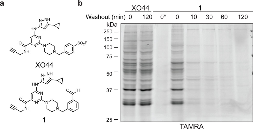
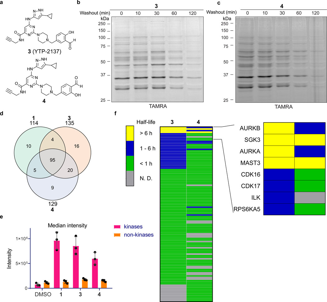
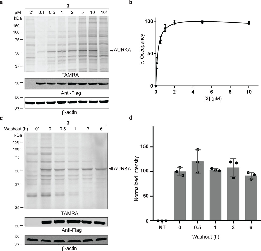
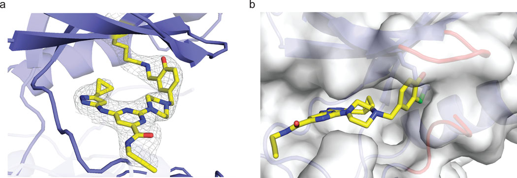
The expansion of the target landscape of covalent inhibitors requires the engagement of nucleophiles beyond cysteine. Although the conserved catalytic lysine in protein kinases is an attractive candidate for a covalent approach, selectivity remains an obvious challenge. Moreover, few covalent inhibitors have been shown to engage the kinase catalytic lysine in animals. We hypothesized that reversible, lysine-targeted inhibitors could provide sustained kinase engagement in vivo, with selectivity driven in part by differences in residence time. By strategically linking benzaldehydes to a promiscuous kinase binding scaffold, we developed chemoproteomic probes that reversibly and covalently engage >200 protein kinases in cells and mice. Probe-kinase residence time was dramatically enhanced by a hydroxyl group ortho to the aldehyde. Remarkably, only a few kinases, including Aurora A, showed sustained, quasi-irreversible occupancy in vivo, the structural basis for which was revealed by X-ray crystallography. We anticipate broad application of salicylaldehyde-based probes to proteins that lack a druggable cysteine.


Conflict of interest statement

Competing interests

T.Y., B.H., P.K., J.R.M., J.C.K., J.D.L., S.N., and J.D.C. are current or former employees of Pfizer. J.T. is a founder of Global Blood Therapeutics, Principia Biopharma, Kezar Life Sciences, Cedilla Therapeutics, and Terremoto Biosciences, and a scientific advisor to Entos.

Figures

Extended Data Fig. 1
Extended Data Fig. 1. Reversible protein labeling by benzaldehyde probe 2.
(a) Chemical structures of 1 and 2. (b) Jurkat cells were treated with 1 or 2 (2 μM, 30 min), followed by compound washout for the indicated times. Cells were lysed in the presence of 25 mM sodium cyanoborohydride, except as indicated (#). After copper-promoted click conjugation with TAMRA-azide, samples were analyzed by in-gel fluorescence and Coomassie blue staining. Data are representative of two independent experiments.
Extended Data Fig. 2
Extended Data Fig. 2. Probe 3 modification of overexpressed SRC.
(a) COS-7 cells were transfected with WT or K295Q (#) Flag-SRC, or not transfected (*), and then treated with the indicated concentrations of probe 3 (30 min). After lysis in the presence of sodium borohydride and TAMRA-azide conjugation, samples were analyzed by in-gel fluorescence and western blotting. Data are representative of two independent experiments. (b) Concentration-dependent labeling of Flag-SRC (n=2, mean values from two independently performed experiments were plotted).
Extended Data Fig. 3
Extended Data Fig. 3. Rapid dissociation of probe 3 from overexpressed SRC.
(a) COS-7 cells were transfected with Flag-SRC, or not transfected (*), and treated with probe 3 (2 μM, 30 min), followed by washout for the indicated times. After lysis in the presence of sodium borohydride and TAMRA-azide conjugation, samples were analyzed by in-gel fluorescence and western blotting. (b) Normalized fluorescence intensity of ~65 kDa band corresponding to Flag-SRC (mean ± SD, n = 3).
Extended Data Fig. 4
Extended Data Fig. 4. Dissociation of probe 3 from recombinant AURKA and SRC.
AURKA or SRC (5 μM) was treated with probe 3 (5.1 μM) in 50 mM HEPES, pH 8.0 at RT for 5 min, followed by 20-fold dilution into buffer containing 10 μM XO44. The percentage of XO44-modified and unmodified kinase was quantified by LC-MS at the indicated time points, and % unmodified kinase (corresponding to probe 3-bound kinase) was plotted vs. time (n = 2, mean values from two independently performed experiments were plotted).
Extended Data Fig. 5
Extended Data Fig. 5. Related to Figure 5.
Volcano plot showing log2 fold-change and significance (−log10 p-value; two tailed t-test assuming unequal variance) of proteins enriched by probe 3 (t=3 h post-dose) vs. vehicle. Red, kinases; gray, non-kinases.
Figure 1.
Figure 1.
Benzaldehyde probe 1 reversibly labels cellular proteins. (a) Chemical structures of XO44 and probe 1. (b) Jurkat cells were treated with XO44 or 1 (2 μM, 30 min), followed by compound washout for the indicated times. Cells treated with 1 were lysed in the presence of 25 mM sodium cyanoborohydride, except as indicated (*). After copper-promoted click conjugation with TAMRA-azide, samples were analyzed by in-gel fluorescence and Coomassie blue staining (Supplementary Figure 1). Data are representative of two independent experiments.
Figure 2.
Figure 2.
Salicylaldehyde probes exhibit prolonged kinase residence time in cells. (a) Salicylaldehyde probes 3 and 4. (b,c) Jurkat cells were treated with 3 (b) or 4 (c) (2 μM, 30 min), followed by compound washout for the indicated times and lysis in the presence of sodium borohydride. After click conjugation with TAMRA-azide, samples were analyzed by in-gel fluorescence and Coomassie blue staining (Supplementary Figure 2). Data are representative of two independent experiments. (d-f) Jurkat cells were treated with DMSO or probes 1, 3, and 4 (2 μM, 30 min), followed by washout as described in the main text. After click conjugation with biotin-azide, modified proteins were enriched with Neutravidin-agarose and digested on-bead with trypsin. Enriched peptides were analyzed by LC-MS/MS and label-free quantitation (LFQ). (d) Shared and unique kinases enriched by each probe. (e) Median intensity values (LFQ) for identified kinases and non-kinases (mean ± SD, n=3). (f) Heat map showing estimated half-lives (t1/2) of kinases enriched from cells treated with probes 3 and 4, followed by washout for 0, 1, 3, and 6 hr. Inset depicts kinases with prolonged engagement post-washout. Grey bars (N.D.): kinase not detected or did not meet criteria for t1/2 determination (see main text).
Figure 3.
Figure 3.
Quasi-irreversible engagement of AURKA in cells. (a) COS-7 cells were transfected with WT or K162Q (#) Flag-AURKA and treated with probe 3 (30 min). After lysis in the presence of sodium borohydride and TAMRA-azide conjugation, samples were analyzed by in-gel fluorescence and western blotting. (b) Concentration-dependent labeling of Flag-AURKA (~50 kDa TAMRA band, mean ± SD, n=3). (c,d) COS-7 cells were transfected with Flag-AURKA and treated with probe 3 (2 μM, 30 min), followed by washout for the indicated times. Samples were analyzed as described above (mean ± SD, n=3). * and NT indicate non-transfected cells.
Figure 4.
Figure 4.
Co-crystal structure of AURKA bound to salicylaldehyde 3. (a) Salicylaldehyde 3 forms an imine bond with the catalytic lysine of AURKA. Electron density map (2Fo-Fc) is shown at a contour level of 1σ. (b) Surface rendering of AURKA bound to probe 3, highlighting the Gly-rich loop (red, top right) and the activation segment (red, bottom right), both of which shield the salicylaldimine from bulk water (imine carbon in green).
Figure 5.
Figure 5.
Quantifying kinase occupancy by salicylaldehyde 3 in mice. (a) Mice were dosed with vehicle or probe 3 (25 or 50 mg/kg) by subcutaneous injection. At 1, 3, and 7 h post-dose, plasma and spleens were collected for quantification of probe 3 concentrations and kinase occupancy, respectively. Spleens from each time point were selected for chemoproteomic analysis (biotin-azide conjugation, streptavidin pulldown, 10-plex TMT quantitation) based on the probe 3 plasma levels quantified from the same mice (see main text and Supplementary Table 2). Spleens from vehicle-treated mice were collected at t=7 h post-injection. (b-c) Plasma concentrations of probe 3 and relative occupancy of the indicated kinases (normalized to t=3 h) are plotted for each time point (t=1 and 7 h, n=2; t=3 h, n=3). (d) Scatter plot comparing relative kinase occupancy (log2 fold-change vs. kinase occupancy at t=3 h) at t=7 h vs. t=1 h. Each circle represents one kinase, with AURKA, AURKB, and SGK3 showing the highest occupancy at t=7 h, despite decreased plasma levels of probe 3.
Figure 6.
Figure 6.
Salicylaldehyde probe 3 reveals PF-06873600 target engagement in vivo. (a) Mice were dosed orally with vehicle (n=5) or PF-06873600 (50 mg/kg, n=5), and all mice were co-dosed with probe 3 (10 mg/kg) by subcutaneous injection. After 1 h, spleens were collected for chemoproteomic analysis. (b) Chemical structure of PF-06873600. (c) Volcano plot showing log2 fold-change and significance (−log10 p-value) between proteins enriched by probe 3 from mice treated with PF-06873600 vs. vehicle. Red, kinases; gray, non-kinases.

References

    1. Honigberg LA; Smith AM; Sirisawad M; Verner E; Loury D; Chang B; Li S; Pan Z; Thamm DH; Miller RA; Buggy JJ The Bruton tyrosine kinase inhibitor PCI-32765 blocks B-cell activation and is efficacious in models of autoimmune disease and B-cell malignancy. Proc Natl Acad Sci U S A 2010, 107 (29), 13075–13080. https://www.pnas.org/content/pnas/107/29/13075.full.pdf - PMC - PubMed
    1. Byrd JC; Furman RR; Coutre SE; Flinn IW; Burger JA; Blum KA; Grant B; Sharman JP; Coleman M; Wierda WG; Jones JA; Zhao W; Heerema NA; Johnson AJ; Sukbuntherng J; Chang BY; Clow F; Hedrick E; Buggy JJ; James DF; O’Brien S. Targeting BTK with Ibrutinib in Relapsed Chronic Lymphocytic Leukemia. N Engl J Med 2013, 369 (1), 32–42. https://www.nejm.org/doi/full/10.1056/NEJMoa1215637 - DOI - PMC - PubMed
    1. Finlay MRV; Anderton M; Ashton S; Ballard P; Bethel PA; Box MR; Bradbury RH; Brown SJ; Butterworth S; Campbell A; Chorley C; Colclough N; Cross DAE; Currie GS; Grist M; Hassall L; Hill GB; James D; James M; Kemmitt P; Klinowska T; Lamont G; Lamont SG; Martin N; McFarland HL; Mellor MJ; Orme JP; Perkins D; Perkins P; Richmond G; Smith P; Ward RA; Waring MJ; Whittaker D; Wells S; Wrigley GL Discovery of a Potent and Selective EGFR Inhibitor (AZD9291) of Both Sensitizing and T790M Resistance Mutations That Spares the Wild Type Form of the Receptor. J Med Chem 2014, 57 (20), 8249–8267. 10.1021/jm500973a - DOI - PubMed
    1. Jänne PA; Yang JC-H; Kim D-W; Planchard D; Ohe Y; Ramalingam SS; Ahn M-J; Kim S-W; Su W-C; Horn L; Haggstrom D; Felip E; Kim J-H; Frewer P; Cantarini M; Brown KH; Dickinson PA; Ghiorghiu S; Ranson M. AZD9291 in EGFR Inhibitor–Resistant Non–Small-Cell Lung Cancer. N Engl J Med 2015, 372 (18), 1689–1699. https://www.nejm.org/doi/full/10.1056/NEJMoa1411817 - DOI - PubMed
    1. Lanman BA; Allen JR; Allen JG; Amegadzie AK; Ashton KS; Booker SK; Chen JJ; Chen N; Frohn MJ; Goodman G; Kopecky DJ; Liu L; Lopez P; Low JD; Ma V; Minatti AE; Nguyen TT; Nishimura N; Pickrell AJ; Reed AB; Shin Y; Siegmund AC; Tamayo NA; Tegley CM; Walton MC; Wang H-L; Wurz RP; Xue M; Yang KC; Achanta P; Bartberger MD; Canon J; Hollis LS; McCarter JD; Mohr C; Rex K; Saiki AY; San Miguel T; Volak LP; Wang KH; Whittington DA; Zech SG; Lipford JR; Cee VJ Discovery of a Covalent Inhibitor of KRASG12C (AMG 510) for the Treatment of Solid Tumors. J Med Chem 2020, 63 (1), 52–65. 10.1021/acs.jmedchem.9b01180 - DOI - PubMed

Publication types