. 2022 May 19;12(1):8489.
doi: 10.1038/s41598-022-12597-4.

Initial TK-deficient HSV-1 infection in the lip alters contralateral lip challenge immune dynamics

Affiliations

Initial TK-deficient HSV-1 infection in the lip alters contralateral lip challenge immune dynamics

Antoine Rousseau et al. Sci Rep. .

Abstract

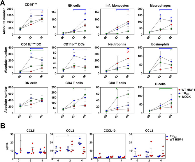
Primary infection with herpes simplex type 1 (HSV-1) occurring around the mouth and nose switches rapidly to lifelong latent infection in sensitive trigeminal ganglia (TG) neurons. Sporadic reactivation of these latent reservoirs later in life is the cause of acute infections of the corneal epithelium, which can cause potentially blinding herpes simplex keratitis (HSK). There is no effective vaccine to protect against HSK, and antiviral drugs provide only partial protection against recurrences. We previously engendered an acute disease-free, non-reactivating latent state in mice when challenged with virulent HSV-1 in orofacial mucosa, by priming with non-neurovirulent HSV-1 (TKdel) before the challenge. Herein, we define the local immune infiltration and inflammatory chemokine production changes after virulent HSV-1 challenge, which were elicited by TKdel prime. Heightened immunosurveillance before virulent challenge, and early enhanced lymphocyte-enriched infiltration of the challenged lip were induced, which corresponded to attenuation of inflammation in the TG and enhanced viral control. Furthermore, classical latent-phase T cell persistence around latent HSV-1 reservoirs were severely reduced. These findings identify the immune processes that are likely to be responsible for establishing non-reactivating latent HSV-1 reservoirs. Stopping reactivation is essential for development of efficient vaccine strategies against HSV-1.


Conflict of interest statement

The authors declare no competing interests.

Figures

Figure 1
Figure 1
Infection regime, WT HSV-1 gene expression, and immune cell infiltration in the iTG. Schematic of the superinfection model, versus the challenge of unprimed (naïve) mice (A). Naïve mice were infected in the lip with either PBS (Mock), WT HSV-1, or with TKdel on day 0. Superinfection model mice were initially infected with TKdel in the right lip on day -4 and then challenged with WT HSV-1 on day 0 in the left lip. On different days for each group, lip and TG were harvested from individual mice, and WT HSV-1 thymidine kinase gene expression was measured from TG by RT-qPCR (B), while CD45+ve cells were defined and absolute numbers enumerated by flow cytometry (C). Each point represents n = 4 individual biological replicates. Colored bars indicate when the color-matched group was significantly different to uninfected day 0 (p < 0.03; permutation test). Red asterisks identify times when WT HSV-1 was significantly different to PBS mock infected (green) and superinfection model. Orange asterisks indicate significant difference between superinfection model versus infection in unprimed mice (permutation test, p < 0.03). A significant difference between mock and only the color-corresponding group is indicated with a colored P. Figures were formulated using Flowjo, Excel and Prism.
Figure 2
Figure 2
Immune subsets in the iTG. Naïve mice were infected in the lip with WT HSV-1, PBS (mock; green line), or with TKdel. Mice of the superinfection model received an initial inoculation of TKdel in the right lip on day -4, and were challenged with WT HSV-1 on day 0 in the left lip. On different days for each group, immune subsets (AK) were defined and absolute numbers quantified by flow cytometry in iTG single cell suspensions. Colored bars indicate significant differences from uninfected tissue at day 0. Red asterisks identify when WT HSV-1 compared to PBS mock infection (green) and superinfection model was significantly different (p < 0.03; permutation test). Colored P indicates significance between Mock and the color-corresponding group. Each point represents n = 4 individual biological replicates. Figures were formulated using Flowjo, Excel and Prism.
Figure 3
Figure 3
Inflammatory chemokines in the iTG. Naïve mice were infected in the lip with TKdel (A), or WT HSV-1 (B) on day 0. Superinfection model mice were initially infected with TKdel in the right lip on day -4, and then challenged with WT HSV-1 in the left lip on day 0 (C). Inflammatory chemokines in non-denatured iTG lysates were quantified using multi-analyte flow cytometry assay. Bars indicate significant difference from uninfected day 0 (p < 0.03; T-test). Each point represents n = 4 independent biological replicates. Figures were formulated using LEGENDplex software, Excel and Prism.
Figure 4
Figure 4
WT HSV-1 gene expression and immune infiltrate at the lip inoculation site. Naïve mice were infected in the lip with PBS (Mock; green), WT HSV-1, or TKdel on day 0. Superinfection model mice were initially inoculated with TKdel in the right lip on day -4 and then superinfected with WT HSV-1 on day 0 in the left lip. On different days for each group, thymidine kinase viral gene expression was measured by RT-qPCR in inoculation site lip tissue (A), while CD45+ve cells were defined and absolute numbers enumerated from single cell suspensions by flow cytometry (B). Colored bars indicate significant differences from uninfected tissue at day 0 (permutation test, p < 0.01), while asterisks indicate significance between both unprimed WT HSV-1 and Mock (green) versus superinfection model (orange), or both superinfection model and PBS mock infection versus unprimed WT HSV-1 (red) (p < 0.03; permutation test). Each point represents n = 4 individual biological replicates. Figures were formulated using Flowjo, Excel and Prism.
Figure 5
Figure 5
Immune subsets at the lip site of inoculation. Naïve mice were infected on day 0 with WT HSV-1, PBS (mock), or TKdel. Mice of the superinfection model received an initial inoculation of TKdel in the right lip on day -4, and were superinfected on day 0 in the left lip with WT HSV-1. On different days in each group, immune subsets (AK) were defined and absolute numbers quantified by flow cytometry from single cell suspensions of the lip site of virus inoculation. Colored bars indicate significance from uninfected tissue on day 0. Red asterisks indicate a significant difference for unprimed WT HSV-1-infected mice compared to PBS mock infection (green) and superinfection model mice, and orange asterisks indicate significance for superinfection model compared to Mock and WT HSV-1, and (p < 0.03; permutation test). Each point represents n = 4 individual biological replicates. Figures were formulated using Flowjo, Excel and Prism.
Figure 6
Figure 6
Inflammatory chemokines at the lip site of inoculation. Naïve mice were infected in the lip with TKdel (A), or WT HSV-1 (B) on day 0. Mice of the superinfection model were initially inoculated with TKdel in the right lip on day -4 and then superinfected with WT HSV-1 on day 0 in the left lip (C). Mice were randomly culled at indicated time points and inflammatory chemokines in non-denatured lip tissue lysates were quantified using multi-analyte flow cytometry assay. Bars indicate significant difference from uninfected day 0 (p < 0.03; T-test). Each point represents n = 4 independent biological replicates. Figures were formulated using LEGENDplex software, Excel and Prism.
Figure 7
Figure 7
Immune subsets and inflammatory chemokines in the lip contralateral to prime. Naïve mice were infected with WT HSV-1, PBS (Mock), or TKdel. On different days for each group, immune subsets were defined and absolute numbers enumerated from single cell suspensions by flow cytometry from lip tissue contralateral to the inoculation site (A), or inflammatory chemokines were quantified in non-denatured lip tissue lysates (B). Colored bars indicating significant difference from uninfected tissue on day 0. Blue asterisk indicates a significant difference for TKdel compared to PBS mock infection (green) and WT HSV-1 (p < 0.03; permutation test). Colored P indicates a significant difference between the color-corresponding group and the Mock group. Black asterisks indicate a significant difference (p < 0.03; T-test). Each point represents n = 4 individual biological replicates. Figures were formulated using Flowjo, Excel and Prism.

Similar articles

Cited by

References

    1. Liesegang TJ. Epidemiology of ocular herpes simplex. Natural history in Rochester. Minn. 1950 through 1982. Arch. Ophthalmol. 1989;107:1160–1165. doi: 10.1001/archopht.1989.01070020226030. - DOI - PubMed
    1. Labetoulle M, et al. Incidence of herpes simplex virus keratitis in France. Ophthalmology. 2005;112:888–895. doi: 10.1016/j.ophtha.2004.11.052. - DOI - PubMed
    1. Young RC, Hodge DO, Liesegang TJ, Baratz KH. Incidence, recurrence, and outcomes of herpes simplex virus eye disease in Olmsted County, Minnesota, 1976–2007: The effect of oral antiviral prophylaxis. Arch. Ophthalmol. 2010;128:1178–1183. doi: 10.1001/archophthalmol.2010.187. - DOI - PMC - PubMed
    1. Reynaud C, et al. Persistent impairment of quality of life in patients with herpes simplex keratitis. Ophthalmology. 2017;124:160–169. doi: 10.1016/j.ophtha.2016.10.001. - DOI - PubMed
    1. HEDS Acyclovir for the prevention of recurrent herpes simplex virus eye disease. New Engl. J. Med. 1998;339:300–306. doi: 10.1056/nejm199807303390503. - DOI - PubMed

Publication types