. 2022 May 19;22(1):140.
doi: 10.1186/s12866-022-02554-6.

Localization of EccA3 at the growing pole in Mycobacterium smegmatis

Affiliations

Localization of EccA3 at the growing pole in Mycobacterium smegmatis

Nastassja L Kriel et al. BMC Microbiol. .

Abstract

Background: Bacteria require specialized secretion systems for the export of molecules into the extracellular space to modify their environment and scavenge for nutrients. The ESX-3 secretion system is required by mycobacteria for iron homeostasis. The ESX-3 operon encodes for one cytoplasmic component (EccA3) and five membrane components (EccB3 - EccE3 and MycP3). In this study we sought to identify the sub-cellular location of EccA3 of the ESX-3 secretion system in mycobacteria.

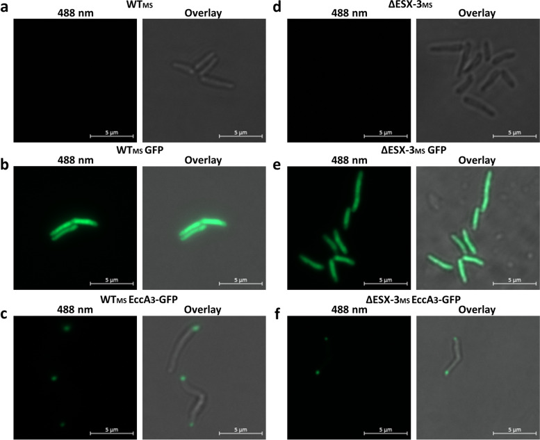
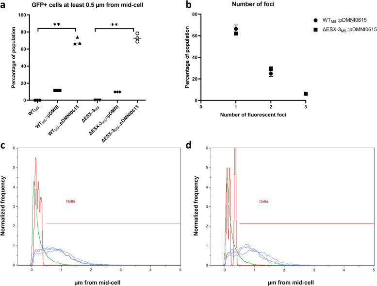
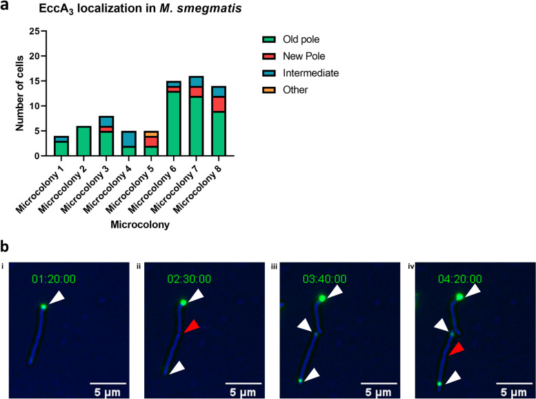
Results: Fluorescently tagged EccA3 localized to a single pole in the majority of Mycobacterium smegmatis cells and time-lapse fluorescent microscopy identified this pole as the growing pole. Deletion of ESX-3 did not prevent polar localization of fluorescently tagged EccA3, suggesting that EccA3 unipolar localization is independent of other ESX-3 components. Affinity purification - mass spectrometry was used to identify EccA3 associated proteins which may contribute to the localization of EccA3 at the growing pole. EccA3 co-purified with fatty acid metabolism proteins (FAS, FadA3, KasA and KasB), mycolic acid synthesis proteins (UmaA, CmaA1), cell division proteins (FtsE and FtsZ), and cell shape and cell cycle proteins (MurS, CwsA and Wag31). Secretion system related proteins Ffh, SecA1, EccA1, and EspI were also identified.

Conclusions: Time-lapse microscopy demonstrated that EccA3 is located at the growing pole in M. smegmatis. The co-purification of EccA3 with proteins known to be required for polar growth, mycolic acid synthesis, the Sec secretion system (SecA1), and the signal recognition particle pathway (Ffh) also suggests that EccA3 is located at the site of active cell growth.

Keywords: ESX-3; EccA3; Mycobacterium; Polar localization; Type VII secretion.


Conflict of interest statement

The authors declare that they have no competing interests.

Figures

Fig. 1
Fig. 1
Unipolar localization of EccA3 in Mycobacterium smegmatis. Fluorescent microscopy demonstrated no GFP fluorescence of WTMS (a) and ΔESX-3MS (d) cells. GFP fluorescence was detected throughout the cell in positive control WTMS::pDMNI (b) and ΔESX-3MS::pDMNI (e) M. smegmatis. EccA3 was found to reside at a single pole within WTMS::pDMNI0615 (c) and ΔESX-3MS::pDMNI0615 (f) cells
Fig. 2
Fig. 2
Localization of EccA3 within a population of Mycobacterium smegmatis. Three independent clonal populations were used for imaging flow cytometry experiments. a Imaging flow cytometry was used to demonstrate that more than 60% of clonal populations of WTMS::pDMNI0615 and ΔESX-3MS::pDMNI0615 had a GFP focus at least 0.5 μm from the mid-cell when compared to no GFP (WTMS and ΔESX-3MS) and GFP positive controls (WTMS::pDMNI and ΔESX-3MS::pDMNI). Unipolar localisation was significant following a Kruskal-Wallis test with a p-value of 0.0036 for both WTMS and ΔESX-3MS (indicated with **). b The majority of GFP positive WTMS::pDMNI0615 and ΔESX-3MS::pDMNI0615 cells with a fluorescent focus at least 0.5 μm from the mid-cell had only one fluorescent focus. c The histogram shows distance of a fluorescent foci from the mid-cell of populations WTMS (red), WTMS::pDMNI (green), WTMS::pDMNI0615.1 (blue), WTMS::pDMNI0615.2 (blue-purple), WTMS::pDMNI0615.3 (purple). d The distance of fluorescent foci from the mid-cell of ΔESX-3MS (red), ΔESX-3MS::pDMNI (green), ΔESX-3MS::pDMNI0615.1 (blue), ΔESX-3MS::pDMNI0615.2 (blue-purple), ΔESX-3MS::pDMNI0615.3 (purple) populations. A population with GFP fluorescence located at least 0.5 μm from the mid-cell, represented by the red line called Delta, was used to score the number of fluorescent foci per cell
Fig. 3
Fig. 3
EccA3 localizes at the growing pole in Mycobacterium smegmatis. a Time-lapse imaging of eight independent clonal populations demonstrated that EccA3 localized at the old pole in 52 of the 73 cells imaged. b A single WTMS::pDMNI0615 cell with a fluorescent focus at the pole (i, white arrow) can be seen dividing into two cells (ii) with a red arrow at the septum. A second fluorescent foci representing EccA3-GFP can be seen at the old pole (iii, white arrow) before a third fluorescent foci (iv, white arrow) is detected following cell division (iv, red arrow at the septum). One of the fluorescent foci detected previously at the old pole (iii, white arrow) migrated to be polar adjacent (iv, white arrow)

References

    1. Gey Van Pittius NC, Gamieldien J, Hide W, Brown GD, Siezen RJ, Beyers AD. The ESAT-6 gene cluster of Mycobacterium tuberculosis and other high G+C Gram-positive bacteria. Genome Biol. 2001;2(10):1–18. doi: 10.1186/gb-2001-2-10-research0044. - DOI - PMC - PubMed
    1. Pallen MJ. The ESAT-6/WXG100 superfamily - And a new Gram-positive secretion system? Trends Microbiol. 2002;10(5):209–212. doi: 10.1016/S0966-842X(02)02345-4. - DOI - PubMed
    1. Renshaw PS, Panagiotidou P, Whelan A, Gordon SV, Glyn Hewinson R, Williamson RA, et al. Conclusive evidence that the major T-cell antigens of the Mycobacterium tuberculosis complex ESAT-6 and CFP-10 form a tight, 1:1 complex and characterization of the structural properties of ESAT-6, CFP-10, and the ESAT-6·CFP-10 complex. Implications for p. J Biol Chem. 2002;277(24):21598–21603. doi: 10.1074/jbc.M201625200. - DOI - PubMed
    1. Fortune SM, Jaeger A, Sarracino DA, Chase MR, Sassetti CM, Sherman DR, et al. Mutually dependent secretion of proteins required for mycobacterial virulence. Proc Natl Acad Sci U S A. 2005;102(30):10676–10681. doi: 10.1073/pnas.0504922102. - DOI - PMC - PubMed
    1. Cole ST, Brosch R, Parkhill J, Garnier T, Churcher C, Harris D, et al. Deciphering the biology of Mycobacterium tuberculosis from the complete genome sequence. Nature. 1998;393(6685):537–544. doi: 10.1038/31159. - DOI - PubMed

Publication types

MeSH terms