. 2022 Apr 25;15(9):3100.
doi: 10.3390/ma15093100.

Electrochemical and In Vitro Biological Evaluation of Bio-Active Coatings Deposited by Magnetron Sputtering onto Biocompatible Mg-0.8Ca Alloy

Affiliations

Electrochemical and In Vitro Biological Evaluation of Bio-Active Coatings Deposited by Magnetron Sputtering onto Biocompatible Mg-0.8Ca Alloy

Ana-Iulia Bița et al. Materials (Basel). .

Abstract

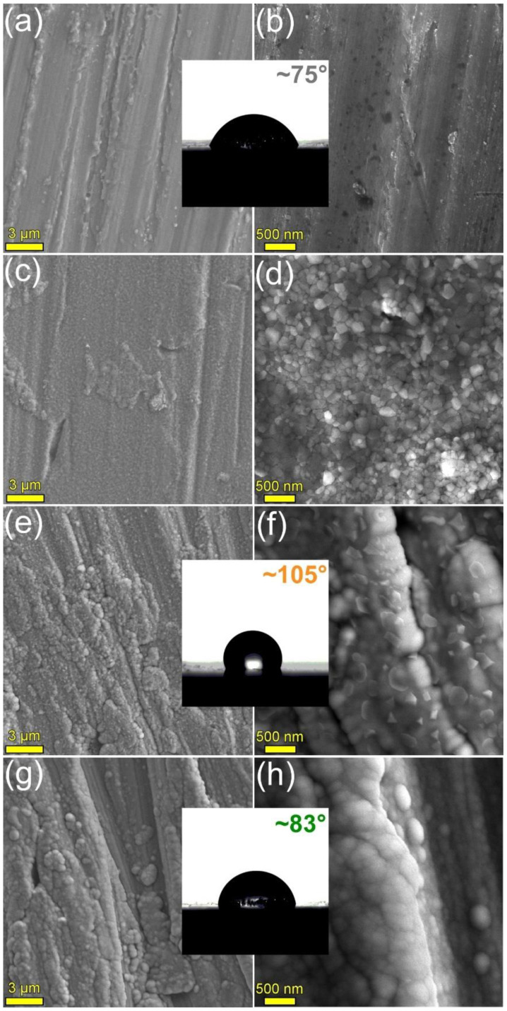
The use of resorbable magnesium alloys in the design of implants represents a new direction in the healthcare domain. Two main research avenues are currently explored for developing or improving metallic biomaterials: (i) increase of their corrosion resistance by designed compositional and structural modifications, and (ii) functionalization of their surfaces by coating with ceramic or polymeric layers. The main objective of this work was to comparatively assess bio-functional coatings (i.e., highly-crystallized hydroxyapatite and silica-rich glass) deposited by radio-frequency magnetron sputtering (RF-MS) on a biodegradable Mg-0.8Ca alloy (0.8 wt.% of Ca). After probing their morphology (by scanning electron microscopy) and structure (by Fourier transform infrared spectroscopy and grazing incidence X-ray diffraction), the corrosion resistance of the RF-MS coated Mg-0.8Ca substrates was electrochemically tested (in synthetic biological media with different degrees of biomimicry), and their cytocompatibility was assessed in osteoblast and fibroblast cell cultures. By collective assessment, the most promising performances, in terms of mass loss (~7% after 12 days), hydrogen release rate (~6 mL/cm2 after 12 days), electrochemical corrosion parameters and cytocompatibility, were obtained for the crystalline HA coating.

Keywords: Mg-0.8Ca alloy; bio-active coatings; bio-glass; hydroxyapatite; magnetron sputtering.


Conflict of interest statement

The authors declare no conflict of interest. The funders had no role in the design of the study; in the collection, analyses, or interpretation of data; in the writing of the manuscript, or in the decision to publish the results.

Figures

Figure 1
Figure 1
Cross-sectional SEM images of the (a) HA2 (post-deposition heat-treated at 500 °C/1 h in air) and (b) BG2 (as-sputtered) films deposited onto Si substrates.
Figure 2
Figure 2
Characteristic FE-HRSEM images for the surface of the (a,b) bare as-fabricated, (c,d) bare 500 °C annealed in air, (e,f) HA2 (post-deposition heat-treated at 500 °C/1 h in air), and (g,h) BG2 (as-sputtered) RF-MS functionalized Mg-0.8Ca substrates. Insets: Typical CA images recorded for the surface of the bare and HA- and BG2-coated Mg-0.8Ca specimens.
Figure 3
Figure 3
XRD diagram (collected in Bragg–Brentano geometry) of the Mg-0.8Ca alloy.
Figure 4
Figure 4
(a) XRD patterns (recorded in grazing incidence mode, α = 2°) and (b) FTIR-ATR spectra of the bare as-fabricated and 500 °C-annealed Mg-0.8Ca substrates and of the HA2- (post-deposition heat-treated at 500 °C/1 h in air) and BG2- (as-sputtered) RF-MS functionalized Mg-0.8Ca substrates. Inset in (a): Zoomed region (rescaled on the intensity Y-axis) allowing to visualize the amorphous hump, specific to the BG2-type film.
Figure 5
Figure 5
Mass loss time evolution in (a) DMEM and (b) SBF testing media of the bare and HA2- and BG2-coated Mg-0.8Ca specimens.
Figure 6
Figure 6
Hydrogen release evolution in (a) DMEM and (b) SBF testing media of the bare and HA2- and BG2-coated Mg-0.8Ca specimens.
Figure 7
Figure 7
Tafel plots of the bare and HA2- and BG2-coated Mg-0.8Ca specimens immersed in (a) DMEM and (b) SBF testing media. * Mg-0.8Ca Tafel plots inserted from [41].
Figure 8
Figure 8
(a,b,d,e,g,h) SEM images and (c,f,i) EDS spectra collected subsequent to the corrosion tests performed in DMEM, on the (ac) bare and (df) HA2- and (gi) BG2-coated Mg-0.8Ca specimens.
Figure 9
Figure 9
(a,b,d,e,g,h) SEM images and (c,f,i) EDS spectra collected subsequent to the corrosion tests performed in SBF, on the (ac) bare and (df) HA2- and (gi) BG2-coated Mg-0.8Ca specimens.
Figure 10
Figure 10
Viability/proliferation of (a,b) osteoblast and (c,d) fibroblast cells cultured for 1, 2, 3, and 7 days in (a,c) 24 h and (b,d) 72 h extracts obtained for the bare and HA2- and BG2-coated Mg-0.8Ca specimens.

References

    1. Chen Y., Dou J.J., Yu H.J., Chen C.Z. Degradable magnesium-based alloys for biomedical applications: The role of critical alloying elements. J. Biomater. Appl. 2019;33:1348–1372. doi: 10.1177/0885328219834656. - DOI - PubMed
    1. Staiger M.P., Pietak A.M., Huadmai J., Dias G. Magnesium and its alloys as orthopedic biomaterials: A review. Biomaterials. 2006;27:1728–1734. doi: 10.1016/j.biomaterials.2005.10.003. - DOI - PubMed
    1. Kamrani S., Fleck C. Biodegradable magnesium alloys as temporary orthopaedic implants: A review. Biometals. 2019;32:185–193. doi: 10.1007/s10534-019-00170-y. - DOI - PubMed
    1. Xu L., Pan F., Yu G., Yang L., Zhang E., Yang K. In Vitro and In Vivo Evaluation of the Surface Bioactivity of a Calcium Phosphate Coated Magnesium Alloy. Biomaterials. 2009;30:1512–1523. doi: 10.1016/j.biomaterials.2008.12.001. - DOI - PubMed
    1. Bita A.-I., Antoniac I., Ciuca I. Potential Use of Mg-Ca Alloys for Orthopedic Applications. U.P.B. Sci. Bull. Ser. B. 2016;78:173–184.

LinkOut - more resources