. 2022 Jun 21;121(12):2449-2460.
doi: 10.1016/j.bpj.2022.05.013. Epub 2022 May 18.

Nanosurfer assay dissects β-cardiac myosin and cardiac myosin-binding protein C interactions

Affiliations

Nanosurfer assay dissects β-cardiac myosin and cardiac myosin-binding protein C interactions

Anja M Touma et al. Biophys J. .

Abstract

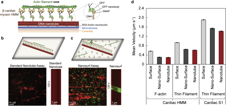
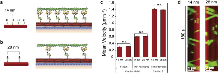
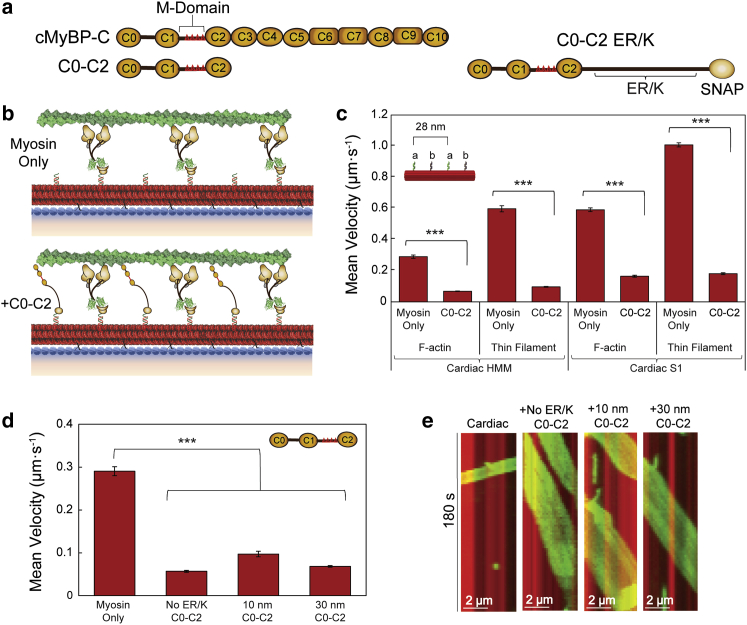
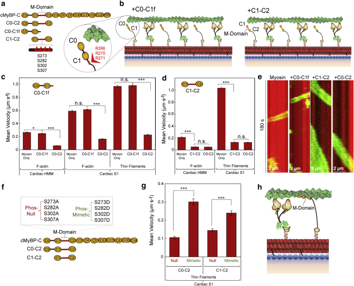
Cardiac myosin-binding protein C (cMyBP-C) modulates cardiac contractility through putative interactions with the myosin S2 tail and/or the thin filament. The relative contribution of these binding-partner interactions to cMyBP-C modulatory function remains unclear. Hence, we developed a "nanosurfer" assay as a model system to interrogate these cMyBP-C binding-partner interactions. Synthetic thick filaments were generated using recombinant human β-cardiac myosin subfragments (HMM or S1) attached to DNA nanotubes, with 14- or 28-nm spacing, corresponding to the 14.3-nm myosin spacing in native thick filaments. The nanosurfer assay consists of DNA nanotubes added to the in vitro motility assay so that myosins on the motility surface effectively deliver thin filaments to the DNA nanotubes, enhancing thin filament gliding probability on the DNA nanotubes. Thin filament velocities on nanotubes with either 14- or 28-nm myosin spacing were no different. We then characterized the effects of cMyBP-C on thin filament motility by alternating HMM and cMyBP-C N-terminal fragments (C0-C2 or C1-C2) on nanotubes every 14 nm. Both C0-C2 and C1-C2 reduced thin filament velocity four- to sixfold relative to HMM alone. Similar inhibition occurred using the myosin S1 construct, which lacks the myosin S2 region proposed to interact with cMyBP-C, suggesting that the cMyBP-C N terminus must interact with other myosin head domains and/or actin to slow thin filament velocity. Thin filament velocity was unaffected by the C0-C1f fragment, which lacks the majority of the M-domain, supporting the importance of this domain for inhibitory interaction(s). A C0-C2 fragment with phospho-mimetic replacement in the M-domain showed markedly less inhibition of thin filament velocity compared with its phospho-null counterpart, highlighting the modulatory role of M-domain phosphorylation on cMyBP-C function. Therefore, the nanosurfer assay provides a platform to precisely manipulate spatially dependent cMyBP-C binding-partner interactions, shedding light on the molecular regulation of β-cardiac myosin contractility.


Conflict of interest statement

Declaration of interests The authors declare no competing interests.

Figures

Figure 1
Figure 1
Optimization of human β-cardiac myosin HMM nanosurfer assay. (a) Schematic of the β-cardiac myosin synthetic thick filament assay with recombinant human β-cardiac myosin HMM (brown) with C-terminal GFP attached to the nanotube (red) via GFP nanobody-SNAP labeled with complementary oligo to the nanotube DNA handle (inset). Nanotubes are attached to the nitrocellulose-coated (blue) coverslip (orange) via BSA-biotin-neutravidin (black Xs). (b) Schematic of standard nanotube assay within a flow cell with motor bound to nanotubes only and nanotubes attached to a coverslip surface blocked with BSA (top). Field of view at 1000× (bottom left) and kymograph (bottom right) for standard nanotube assay (nanotubes in red) with β-cardiac myosin HMM showing the low landing rate of actin filaments (green) on the nanotube surface. (c) Schematic (top) of nanosurfer assay with motor bound to both the nanotubes and the coverslip surface and actin (green) can be observed gliding from the coverslip surface onto the nanotube (green arrow). Field of view at 1000× (bottom left) and kymograph (bottom right) for nanosurfer assay with β-cardiac myosin HMM showing a higher landing rate for actin filaments on the nanotube surface. (d) Velocities of F-actin (left) and regulated thin filaments (middle) with β-cardiac myosin HMM and regulated thin filaments with β-cardiac myosin S1 (right) on a standard in vitro motility surface (light gray), on the coverslip surface in the nanosurfer assay (nano-surface; dark gray), and on the nanotubes in the nanosurfer assay (red). Mean velocities represented as μm·s−1 ± SE. N = 132–390 filaments from three to five independent protein preparations per condition.
Figure 2
Figure 2
Motor spacing does not affect β-cardiac HMM motility. (a and b) Schematic of the synthetic thick filament with nanotube motor spacing of 14 nm (a) or 28 nm (b). (c) Velocities of F-actin (left) and regulated thin filaments (middle) on nanotubes decorated with β-cardiac myosin HMM or regulated thin filaments on nanotubes decorated with β-cardiac myosin S1 (right) at 14- or 28-nm spacing in the nanosurfer assay. (d) Kymographs of F-actin filaments (green) traveling on a nanotube (red) with β-cardiac myosin HMM at 14-nm (left) or 28-nm (right) spacing. Mean velocities represented as μm·s−1 ± SE. N = 143–270 filaments from three to five independent protein preparations per condition. Comparisons were performed using Student's t-test. n.s., not significant
Figure 3
Figure 3
C0–C2 inhibits β-cardiac HMM and S1 nanotube motility. (a) Schematic of cMyBP-C domains C0–C10 with the M-domain (red triangles) containing four phosphorylatable serines in the linker region between the C1 and C2 domains and the N-terminal fragment used, C0–C2. C0–C2 schematic (right) shown with the encoded ER/K linker and SNAP tag. (b) Schematics of synthetic thick filaments with β-cardiac myosin HMM bound to oligo a′ at 28-nm intervals (myosin only, top) and interdigitated C0–C2 containing M-domain, bound to oligo b′ (bottom). (c) Velocities of F-actin and regulated thin filaments on nanotubes decorated with β-cardiac myosin HMM (left) or β-cardiac myosin S1 (right) bound to oligo a′ alone versus myosin + C0–C2 containing a 30-nm ER/K bound to oligo b′ in the pattern shown (inset). (d) Mean velocities of F-actin filaments on nanotubes decorated with β-cardiac myosin HMM only versus β-cardiac HMM nanotubes with interdigitated C0–C2 (inset) containing no ER/K helix, a 10-nm ER/K helix, or a 30-nm ER/K helix. (e) Kymographs of F-actin filaments (green) traveling on a nanotube (red) with β-cardiac myosin HMM only versus cardiac nanotubes with interdigitating C0–C2 containing no ER/K helix, a 10-nm ER/K helix, or a 30-nm ER/K helix. Mean velocities represented as μm·s−1 ± SE. N = 82–514 filaments from three to eight independent protein preparations per condition. Significance calculated using Student's t-test or one-way ANOVA where appropriate. Significance is denoted as ∗∗∗p ≤ 0.001.
Figure 4
Figure 4
cMyBP-C M-domain essential for inhibition of β-cardiac myosin HMM and S1 nanotube motility. (a) Schematic of cMyBP-C domains C0–C10 containing the M-domain (red triangles) in the linker region between the C1 and C2 domains and the N-terminal fragments used, including C0–C2, C0–C1f, and C1–C2. The four phosphorylatable serines in the M-domain are represented by red triangles (S273, S282, S302, S307). (b) Schematics of synthetic thick filaments with β-cardiac myosin HMM bound to oligo a′ at 28-nm intervals and interdigitated C0–C1f (left) or C1–C2 (right) containing the entire M-domain, bound to oligo b′. C0–C1f (inset) contains the first 17 amino acids of the M-domain including several arginine residues (R266, R270, R271) oriented away from the actin filament. (c) Velocities of F-actin on nanotubes decorated with β-cardiac myosin HMM (left) and either F-actin (middle) or regulated thin filaments (right) on nanotubes decorated with β-cardiac myosin S1. β-Cardiac nanotubes were either labeled with the motor alone or interdigitated with C0–C1f or C0–C2 containing 30-nm ER/K helices. (d) Velocities of F-actin on nanotubes decorated with β-cardiac myosin HMM (left) and regulated thin filaments on nanotubes decorated with β-cardiac myosin S1 (right) versus β-cardiac nanotubes with interdigitated C1–C2 or C0–C2 containing 30-nm ER/K helices. (e) Kymographs of F-actin filaments (green) traveling on a nanotube (red) with β-cardiac myosin HMM alone versus cardiac nanotubes with interdigitating C0–C1f, C1–C2, or C0–C2. (f) Schematic of cMyBP-C domains C0–C10 with the M-domain (red triangles) containing four phosphorylatable serines in the linker region between the C1 and C2 domains mutated to alanines (phospho-null) or aspartic acids (phospho-mimetic) within the C0–C2 and C1–C2 N-terminal fragments. (g) Mean nanotube velocities of regulated thin filaments on nanotubes decorated with β-cardiac myosin S1 and interdigitating phospho-null or phospho-mimetic C0–C2 (left) or C1–C2 (right) containing 30-nm ER/K helices. (h) Schematic of β-cardiac myosin (brown) synthetic thick filament with cMyBP-C N-terminal fragment (yellow) bound to actin (green), functioning as a tether between thick and thin filaments and reducing velocity. Mean velocities represented as μm·s−1 ± SE. N = 90–410 filaments from three to four independent protein preparations per condition. Significance was calculated using Student's t-test. Significance is denoted as p ≤ 0.05, ∗∗∗p ≤ 0.001.

References

    1. Previs M.J., Previs S.B., et al. Warshaw D.M. Molecular mechanics of cardiac myosin binding protein-C in native thick filaments. Science. 2012;337:1215–1218. doi: 10.1126/science.1223602. - DOI - PMC - PubMed
    1. Moss R.L., Fitzsimons D.P., Ralphe J.C. Cardiac MyBP-C regulates the rate and force of contraction in mammalian myocardium. Circ. Res. 2015;116:183–192. doi: 10.1161/circresaha.116.300561. - DOI - PMC - PubMed
    1. Harris S.P., Lyons R.G., Bezold K.L. In the thick of it: HCM-causing mutations in myosin binding proteins of the thick filament. Circ. Res. 2011;108:751–764. doi: 10.1161/circresaha.110.231670. - DOI - PMC - PubMed
    1. Starr R., Offer G. Polypeptide chains of intermediate molecular weight in myosin preparations. FEBS Lett. 1971;15:40–44. doi: 10.1016/0014-5793(71)80075-3. - DOI - PubMed
    1. Offer G., Moos C., Starr R. A new protein of the thick filaments of vertebrate skeletal myofibrils. J. Mol. Biol. 1973;74:653–676. doi: 10.1016/0022-2836(73)90055-7. - DOI - PubMed

Publication types

LinkOut - more resources