

Naringin in the repair of knee cartilage injury via the TGF-β/ALK5/Smad2/3 signal transduction pathway combined with an acellular dermal matrix
- PMID: 35591936
- PMCID: PMC9072805
- DOI: 10.1016/j.jot.2021.06.004
Naringin in the repair of knee cartilage injury via the TGF-β/ALK5/Smad2/3 signal transduction pathway combined with an acellular dermal matrix
Abstract
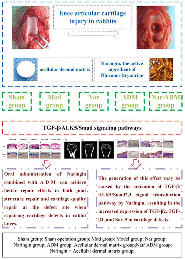
Objective: Based on the expression changes in the TGF-β/ALK5/Smad2/3 signal transduction pathway, the repair of cartilage injury in the rabbit knee joint was investigated and evaluated by oral administration of naringin in combination with acellular dermal matrix implantation.
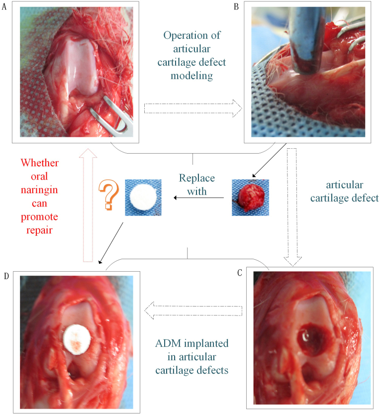
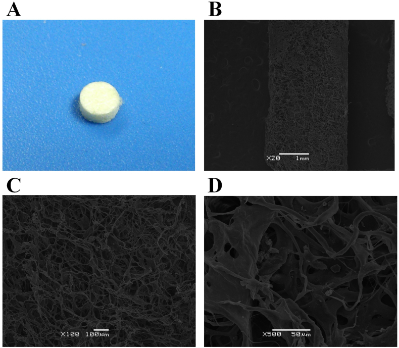
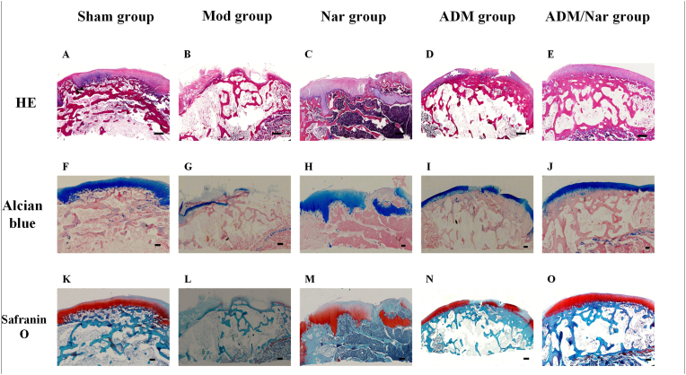
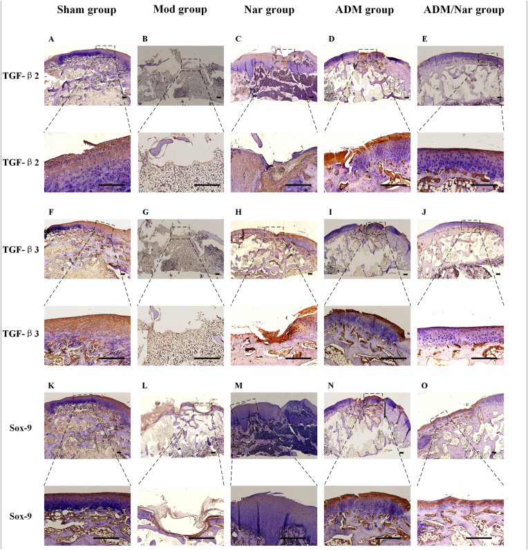
Methods: First, twenty New Zealand white rabbits were randomly divided into five groups: a sham operation group (Sham group), a model group (Mod group), a naringin group (Nar group), an acellular dermal matrix group (ADM group), a naringin + acellular dermal matrix group (Nar/ADM group). After the 12th week, the repaired tissues were assessed for histomorphology and repair content of the repaired site by observing the morphological characteristics of articular cartilage. The International Cartilage Repair Society (ICRS)'s macroscopic evaluation of the cartilage repair scale and the quantitative scoring repair effect of the modified O'Driscoll grading system were used as evaluation criteria. In addition, the structure of the rabbit knee joint was evaluated by micro-CT scan, histological staining (H & E staining, Alcian blue staining, Safranin-O staining) and immunohistochemical staining (TGF-β2 immunostaining, TGF-β3 immunostaining, Sox-9 immunostaining).
Results: ① The observation of the repair morphology of joint defect tissues showed that the repair effects of the Nar and ADM groups were better than that of the Mod group, and the repair effect of Nar/ADM group was the best (P < 0.05). ② Quantitative scoring of joint defect tissue showed that the Nar/ADM group had the best repair efficacy in the quantitative scores of the above two scales compared with the other groups (P < 0.05). ③ Micro-CT scan showed that the ADM group had obvious repair of the defect structure, while the ADM/Nar group had blurred repair boundaries, and the layers of cartilage and subchondral bone were clear. ④ Histological staining (H & E staining, Alcian blue stain, Safranin-O staining) showed that the ADM group had a better effect on the repair of joint structure at the joint defect, the Nar group had a better effect on the repair of cartilage quality at the joint defect, and the ADM/Nar group had satisfactory results in both of the above aspects. ⑤ Immunohistochemical staining (TGF-β2 immunostaining, TGF-β3 immunostaining, Sox-9 immunostaining) revealed that the Nar group showed more abundant expression of the above proteins in articular cartilage defects than the Mod and ADM groups and that the Nar/ADM groups showed extensive TGF-β2, TGF-β3 and Sox-9 protein expression, with uniform expression and smooth distribution.
Conclusions: Oral administration of naringin, the active ingredient of Rhizoma Drynariae, combined with acellular dermal matrix can achieve better repair effects in both joint structure repair and cartilage quality repair at the defect site when repairing cartilage defects in rabbit knees, and the generation of this effect may be caused by the activation of the TGF-β/ALK5/Smad2/3 signal transduction pathway by naringin, resulting in the increased expression of TGF-β2, TGF-β3, and Sox-9 in cartilage defects.
The translational potential of this article: Naringin combined with acellular dermal matrix can facilitate the repair of osteochondral defects and has potential for application in osteochondral tissue engineering.
Keywords: Acellular dermal matrix; Cartilage defect; Nar group Naringin group, ADM group Acellular dermal matrix group; Nar/ ADM group Naringin + Acellular dermal matrix group, ICRS International Cartilage Repair Society; Naringin; Repair; Sham group sham operation group, Mod group model group; Transforming growth factor-β.
© 2021 The Authors.
Conflict of interest statement
The authors have no conflicts of interest to disclose in relation to this article.
Figures


References
-
- Martel-Pelletier J., Barr A.J., Cicuttini F.M., Conaghan P.G., Cooper C., Goldring M.B., et al. Osteoarthritis. Nat Rev Dis Primers. 2016;2:16072. - PubMed
-
- Armiento A.R., Stoddart M.J., Alini M., Eglin D. Biomaterials for articular cartilage tissue engineering: learning from biology. Acta Biomater. 2018;65:1–20. - PubMed
-
- Ma A., Jiang L., Song L., Hu Y., Dun H., Daloze P., et al. Reconstruction of cartilage with clonal mesenchymal stem cell-acellular dermal matrix in cartilage defect model in nonhuman primates. Int Immunopharm. 2013;16(3):399–408. - PubMed
LinkOut - more resources
Full Text Sources
Research Materials
Miscellaneous
