

Evolutionary course of the femoral head osteonecrosis: Histopathological - radiologic characteristics and clinical staging systems
- PMID: 35591937
- PMCID: PMC9072800
- DOI: 10.1016/j.jot.2021.07.004
Evolutionary course of the femoral head osteonecrosis: Histopathological - radiologic characteristics and clinical staging systems
Abstract
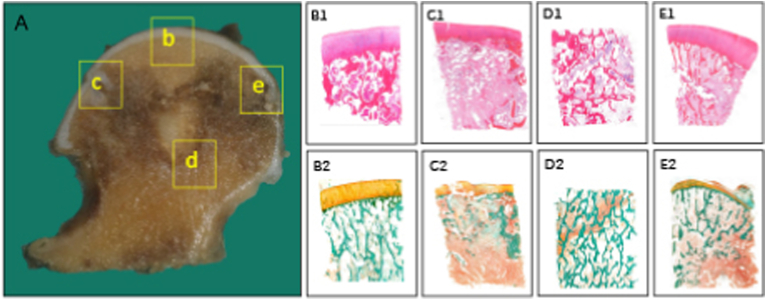
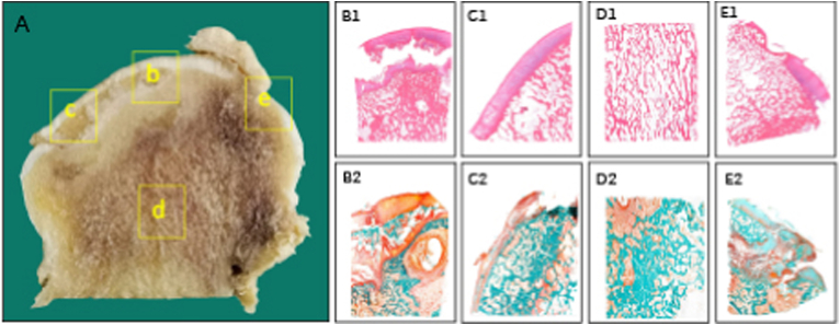
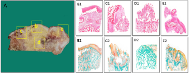
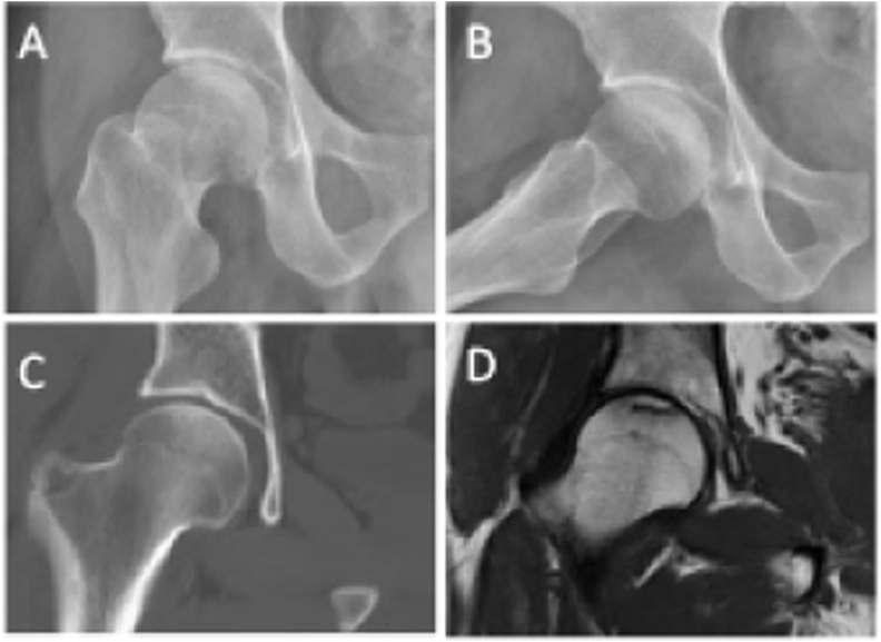
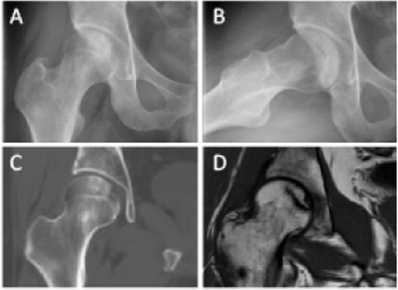
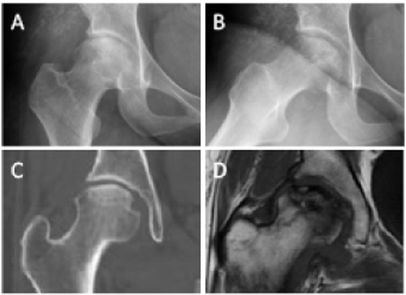
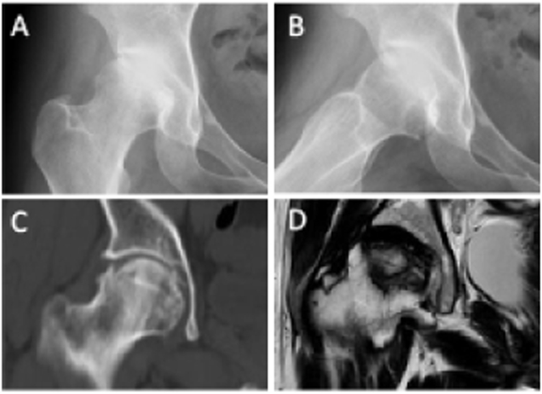
Osteonecrosis of the femoral head (ONFH) is a recalcitrant ischemic disorder, which could be classified into two major categories: traumatic and nontraumatic. Regardless of different risk factors, it has been testified that ONFH results from primitive vascular problems, leading to temporary or permanent loss of blood supply to bone tissue. Histopathological and microarchitectural alterations ensues, which is a gradual evolutionary process involving bone marrow and osteocyte necrosis, progressive destruction of subchondral bone, unsuccessful reparative process, and eventual articular collapse and degenerative arthritis. Based on the imaging features of ONFH, different classification systems have been developed to evaluate the severity and prognosis of the disease, which is pivotal for implementation of treatment strategy, especially the joint-preserving surgery. However, patients classified with the same severity stage, especially in the peri-collapse stage, sometimes responded differently after similar joint-preserving surgery. The unusual phenomenon may be attributed to the limitation of the current imaging classification systems, which might underestimate the disease severity, especially when referring to the early stages. In this review, we briefly summarize the etiology and pathogenesis of ONFH. The imaging features and staging classification systems of ONFH are also described. More importantly, we focus on histopathological and microstructural alterations of the femoral head, and provide an overview of their essential contribution to ONFH progression. Given the observation of discordance between imaging characteristics and histopathological alterations, a substantial amount of research on the relationship between imaging and histopathological features is required to further modify and revise the current wide-accepted classification systems.
Keywords: Histopathology; Imaging; Microarchitecture; Osteonecrosis of the femoral head; Pathogenesis; Staging.
© 2021 The Authors.
Conflict of interest statement
The authors have no conflicts of interest to disclose in relation to this article.
Figures
Similar articles
-
[Clinical significance of different imaging manifestations of osteonecrosis of femoral head in the peri-collapse stage].Zhongguo Xiu Fu Chong Jian Wai Ke Za Zhi. 2021 Sep 15;35(9):1105-1110. doi: 10.7507/1002-1892.202103221. Zhongguo Xiu Fu Chong Jian Wai Ke Za Zhi. 2021. PMID: 34523274 Free PMC article. Chinese.
-
Histopathological aspects and staging systems in non-traumatic femoral head osteonecrosis: an overview of the literature.Acta Biomed. 2016 Apr 15;87 Suppl 1:15-24. Acta Biomed. 2016. PMID: 27104316 Review.
-
Less sclerotic microarchitecture pattern with increased bone resorption in glucocorticoid-associated osteonecrosis of femoral head as compared to alcohol-associated osteonecrosis of femoral head.Front Endocrinol (Lausanne). 2023 Mar 8;14:1133674. doi: 10.3389/fendo.2023.1133674. eCollection 2023. Front Endocrinol (Lausanne). 2023. PMID: 36967755 Free PMC article.
-
Hip preserving surgery with concentrated autologous bone marrow aspirate transplantation for the treatment of asymptomatic osteonecrosis of the femoral head: retrospective review of clinical and radiological outcomes at 6 years postoperatively.BMC Musculoskelet Disord. 2017 Jul 6;18(1):292. doi: 10.1186/s12891-017-1652-8. BMC Musculoskelet Disord. 2017. PMID: 28683795 Free PMC article.
-
Guidelines for clinical diagnosis and treatment of osteonecrosis of the femoral head in adults (2019 version).J Orthop Translat. 2020 Jan 6;21:100-110. doi: 10.1016/j.jot.2019.12.004. eCollection 2020 Mar. J Orthop Translat. 2020. PMID: 32309135 Free PMC article. Review.
Cited by
-
Comparison of outcomes after total hip arthroplasty between patients with osteonecrosis of the femoral head in Association Research Circulation Osseous stage III and stage IV: a five-year follow-up study.J Orthop Surg Res. 2024 Mar 6;19(1):170. doi: 10.1186/s13018-024-04617-y. J Orthop Surg Res. 2024. PMID: 38449033 Free PMC article.
-
Enhanced osteogenic and angiogenic capabilities of adipose-derived stem cells in fish collagen scaffolds for treatment of femoral head osteonecrosis.Sci Rep. 2025 May 26;15(1):18300. doi: 10.1038/s41598-025-03015-6. Sci Rep. 2025. PMID: 40419685 Free PMC article.
-
Advances in experimental models of osteonecrosis of the femoral head.J Orthop Translat. 2023 Feb 3;39:88-99. doi: 10.1016/j.jot.2023.01.003. eCollection 2023 Mar. J Orthop Translat. 2023. PMID: 36819298 Free PMC article. Review.
-
Fibroblast IRF7-mediated chondrocyte apoptosis affects the progression of collapse in steroid-induced osteonecrosis of the femoral head.J Orthop Surg Res. 2025 Mar 18;20(1):292. doi: 10.1186/s13018-025-05557-x. J Orthop Surg Res. 2025. PMID: 40102965 Free PMC article.
-
Bioinformatics and Gene Expression Omnibus Analysis of Key Candidate Genes and Pathways Associated with Femoral Head Necrosis.Iran J Pharm Res. 2024 Jun 2;23(1):e145223. doi: 10.5812/ijpr-145223. eCollection 2024 Jan-Dec. Iran J Pharm Res. 2024. PMID: 39830663 Free PMC article.
References
Publication types
LinkOut - more resources
Full Text Sources
