. 2022 May 12:15:2925-2937.
doi: 10.2147/JIR.S357866. eCollection 2022.

Toll-like Interleukin -1 Receptor Regulator (TILRR) Protein, a Major Modulator of Inflammation, is Expressed in Normal Human and Macaque Tissues and PBMCs

Affiliations

Toll-like Interleukin -1 Receptor Regulator (TILRR) Protein, a Major Modulator of Inflammation, is Expressed in Normal Human and Macaque Tissues and PBMCs

Mohammad Abul Kashem et al. J Inflamm Res. .

Abstract

Purpose: TILRR is a modulator of genes in the NF-κB inflammation pathway. It regulates inflammation-responsive genes, the secretion of inflammatory mediators, and the migration of immune cells. Because inflammation drives the pathogenesis of many infectious and inflammatory diseases, it is important to know the expression of TILRR protein in tissues and cells. This study examined TILRR protein expression in healthy adult human and macaques' tissues and PBMCs (peripheral blood mononuclear cells).

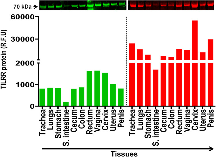
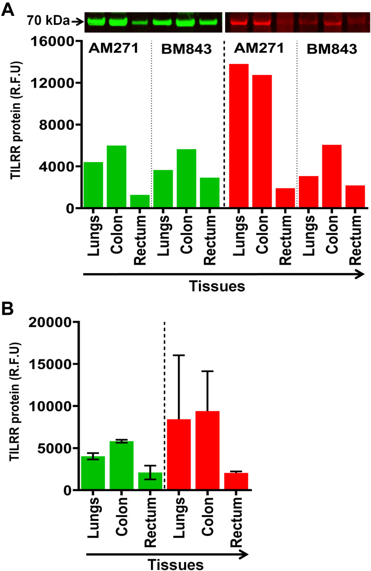
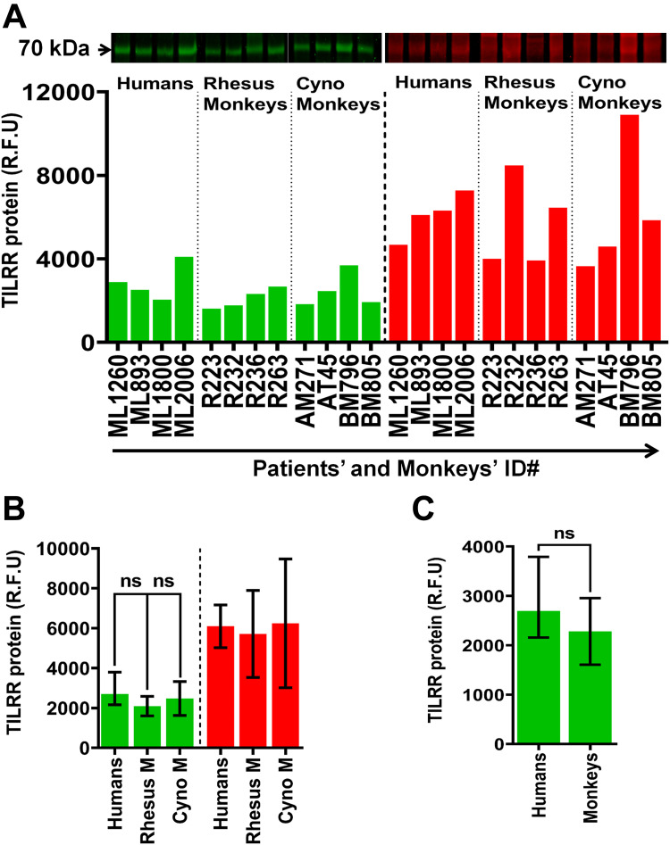
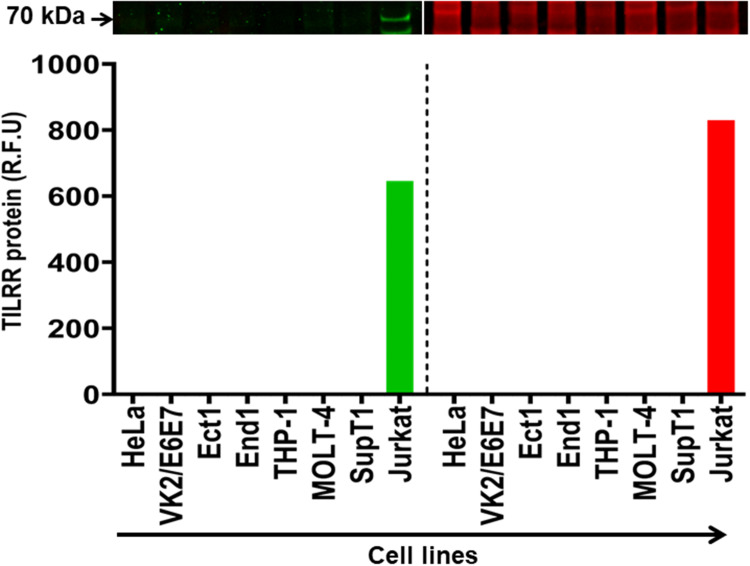
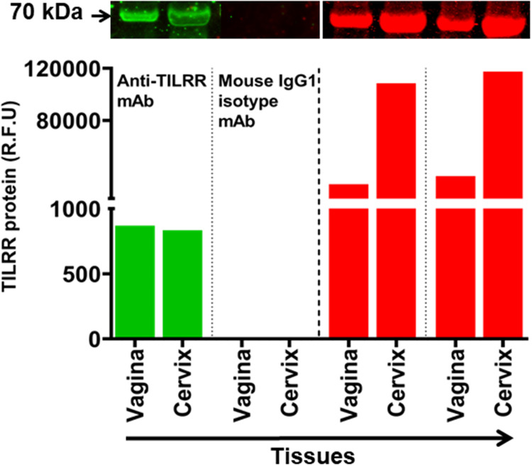
Methods and results: Tissues (trachea, lungs, stomach, small intestine [ileum], cecum, colon, rectum, vagina, cervix, uterus, and penis) and PBMCs from humans and macaques were lysed in RIPA (radioimmunoprecipitation assay) lysis buffer. The TILRR protein was examined by fluorescent Western blot analysis. The relative fluorescence units (rfu) of TILRR protein expression were quantified by Image Studio software (LI-COR). The results showed that adult healthy female (n=1) rectal and cervicovaginal tissues expressed a higher level of TILRR protein than the other tissues (trachea, lungs, stomach, small intestine [ileum], cecum, colon, uterus, and penis) examined. Like humans, the lungs, colon, and rectal tissues of healthy adult female cynomolgus monkeys (Macaca fascicularis) (n=2) expressed the TILRR protein. In addition, PBMCs of healthy adult women (n=4), adult female cynomolgus monkeys (Macaca fascicularis) (n=4), and adult male and female rhesus monkeys (Macaca mulatta) (n=4) showed a similar expression level of TILRR protein (p= 0.2858). TILRR protein was not detected in most of the human cell lines examined, except in Jurkat cells.

Conclusion: Our study for the first time showed that TILRR protein is expressed in healthy adult human and monkey tissues and PBMCs. The TILRR protein in these tissues and PBMCs may play a role in the inflammatory response of these tissues and cells in response to infectious pathogens.

Keywords: PBMCs; TILRR; cynomolgus monkey (Macaca fascicularis); human; inflammation; rhesus monkey (Macaca mulatta); tissues.


Conflict of interest statement

The authors declare no competing interest.

Figures

Figure 1
Figure 1
TILRR protein expression in normal human tissues. Tissue lysates (2.5 µg/lane protein) were examined by Western blot (green color, left) and Coomassie blue staining (red color, right). TILRR protein was probed by primary mouse anti-TILRR F218G4 mAb followed by secondary goat anti-mouse IRDye 800CW antibody (1:10,000; LI-COR). TILRR protein bands (70 kDa, green color, cropped) are shown on the top of the bar graph (see Figure S1A for un-cropped blot image). Untransferred proteins in the gel after iBlot transfer were stained with Coomassie blue staining (G-250, Bio-Rad) and the protein bands (~70 kDa, red color, cropped) are shown on the top of the bar graph (see Figure S1B for un-cropped blot image). The bar graph shows the TILRR protein intensity (R.F.U) for each tissue lysate (n=1) where X-axis demonstrates the normal human tissue lysates and Y-axis indicates the signal intensity (R.F.U). kDa, kiloDalton; RFU, relative fluorescence units; S. intestine, small intestine (ileum).
Figure 2
Figure 2
TILRR protein expression in normal Cynomolgus monkey tissues. (A) Tissue lysates (5.0 µg/lane proteins) were examined by Western blot (green color, left) and Coomassie blue staining (red color, right). TILRR protein was probed by primary mouse anti-TILRR F218G4 mAb followed by secondary goat anti-mouse IRDye 800CW antibody (1:10,000; LI-COR). TILRR protein bands (70 kDa, green color, cropped) are shown on the top of the bar graph (Figure S1C shows the un-cropped blot image). Untransferred proteins in the gel after iBlot transfer were stained with Coomassie blue staining (G-250, Bio-Rad) and the protein bands (~70 kDa, red color, cropped) are shown on the top of the bar graph (Figure S1D shows the un-cropped gel). The bar graph indicates the TILRR protein intensity (R.F.U) for each tissue lysate (n=1). (B) TILRR protein intensity (R.F.U) (median with interquartile range [IQR]) of each tissue cell lysate observed in two cynomolgus monkeys (n=2). The X-axis represents the normal Cynomolgus monkey tissue cell lysates and Y-axis shows the signal intensity (R.F.U). AM271 and BM843 are the monkeys’ identification numbers. kDa, kiloDalton; RFU, relative fluorescence units; n= number of subjects.
Figure 3
Figure 3
TILRR protein expression in normal human and macaque PBMCs. (A) PBMC lysates (14.0 µg/lane proteins) of humans (n=4), rhesus monkeys (n=4), and cynomolgus monkeys (n=4) were analyzed by Western blot (green color, left) and Coomassie blue staining (red color, right). TILRR protein was probed by primary mouse anti-TILRR F218G4 mAb followed by secondary goat anti-mouse IRDye 800CW antibody (1:10,000; LI-COR). TILRR protein bands (70 kDa, green color, cropped) are shown on the top of the bar graph (see Figure S2A for un-cropped blot image). Untransferred proteins in the gel after iBlot transfer were stained with Coomassie blue staining (G-250, Bio-Rad), and the protein bands (~70 kDa, red color, cropped) are shown on the top of the bar graph (see Figure S2B for un-cropped gel). The bar graph shows the TILRR protein intensity (R.F.U). (B) TILRR protein intensity (R.F.U) (median with IQR) of PBMC lysates observed in four study subjects (n=4) including humans, rhesus monkeys, and cynomolgus monkeys. (C) TILRR protein expression between humans and monkeys. Data are shown as median with IQR in figures B and C. A t-test with 95% CI was used for statistical comparisons and all p<0.05 were reported as statistically significant and presented with asterisks. The X-axis of figure A indicates the patients’ and monkeys’ identification numbers and Y-axis shows the signal intensity (R.F.U). kDa, kiloDalton; RFU, relative fluorescence units; n= number of subjects; ID#, identification number, Rhesus M, rhesus monkey; Cyno M, cynomolgus monkey.
Figure 4
Figure 4
TILRR protein expression in human cell lines. Cell line lysates (7 µg/lane protein) were examined by Western blot (green color, left) and Coomassie blue (red color, right). TILRR protein was probed by primary mouse anti-TILRR F218G4 mAb followed by secondary goat anti-mouse IRDye 800CW antibody (1:10,000; LI-COR). TILRR protein bands (70 kDa, green color, cropped) are shown on the top of the bar graph (Figure S3A shows the un-cropped blot image). Untransferred proteins in the gel after iBlot transfer were stained with Coomassie blue staining (G-250, Bio-Rad) and the protein bands (~70 kDa, red color, cropped) are shown on the top of the bar graph (Figure S3B shows the un-cropped gel). The bar graph shows the TILRR protein intensity (R.F.U) for each tissue lysate (n=1) where X-axis demonstrates human cell lines and Y-axis represents signal intensity (R.F.U). kDa, kiloDalton; RFU, relative fluorescence units.
Figure 5
Figure 5
Anti-TILRR mAb is specific for TILRR protein. Tissues lysates (2.5 µg/lane protein) (n=2) were examined by Western blot (green color, left) and Coomassie blue staining (red color, right). TILRR protein was probed by either primary mouse anti-TILRR F218G4 mAb or primary mouse IgG1 mAb (isotype control) (Abcam, Canada) followed by secondary goat anti-mouse IRDye 800CW antibody (1:10,000; LI-COR). TILRR protein bands (70 kDa, green color, cropped) are shown on the top of the bar graph (see Figure S4A for un-cropped blot image). Untransferred proteins in the gel after iBlot transfer were stained with Coomassie blue staining (G-250, Bio-Rad), and the protein bands (~70 kDa, red color, cropped) are shown on the top of the bar graph (see Figure S4B for un-cropped gel). The bar graph shows the TILRR protein intensity (R.F.U) for each tissue lysate where X-axis represents human tissue lysates and Y-axis indicates the signal intensity (R.F.U). kDa, kiloDalton; RFU, relative fluorescence units.

Similar articles

References

    1. Zhang X, Shephard F, Kim HB, et al. TILRR, a Novel IL-1RI Co-receptor, Potentiates MyD88 Recruitment to Control Ras-dependent Amplification of NF-κB. J Biol Chem. 2010;285(10):7222–7232. doi:10.1074/jbc.M109.073429 - DOI - PMC - PubMed
    1. Kashem MA, Li H, Liu LR, et al. The Potential Role of FREM1 and Its Isoform TILRR in HIV-1 Acquisition through Mediating Inflammation. Int J Mol Sci. 2021;22(15):54. - PMC - PubMed
    1. Mac Gabhann F. TILRR Steers Interleukin-1 Signaling: co-Receptor Provides Context and a Therapeutic Target. JACC Basic Transl Sci. 2017;2(4):415–417. - PMC - PubMed
    1. Smith SA, Samokhin AO, Alfadi M, et al. The IL-1RI Co-Receptor TILRR (FREM1 Isoform 2) Controls Aberrant Inflammatory Responses and Development of Vascular Disease. JACC Basic Transl Sci. 2017;2(4):398–414. - PMC - PubMed
    1. Kashem MA, Li H, Toledo NP, et al. Toll-like Interleukin 1 Receptor Regulator Is an Important Modulator of Inflammation Responsive Genes. Front Immunol. 2019;10(272):1–16. - PMC - PubMed