[Preprint]. 2022 May 11:2022.04.28.489942.
doi: 10.1101/2022.04.28.489942.

Immediate myeloid depot for SARS-CoV-2 in the human lung

Affiliations

Immediate myeloid depot for SARS-CoV-2 in the human lung

Mélia Magnen et al. bioRxiv. .

Update in

  • Immediate myeloid depot for SARS-CoV-2 in the human lung.
    Magnen M, You R, Rao AA, Davis RT, Rodriguez L, Bernard O, Simoneau CR, Hysenaj L, Hu KH, Maishan M, Conrad C, Gbenedio OM, Samad B, Consortium TUC, Love C, Woodruff PG, Erle DJ, Hendrickson CM, Calfee CS, Matthay MA, Roose JP, Sil A, Ott M, Langelier CR, Krummel MF, Looney MR. Magnen M, et al. Sci Adv. 2024 Aug 2;10(31):eadm8836. doi: 10.1126/sciadv.adm8836. Epub 2024 Jul 31. Sci Adv. 2024. PMID: 39083602 Free PMC article.

Abstract

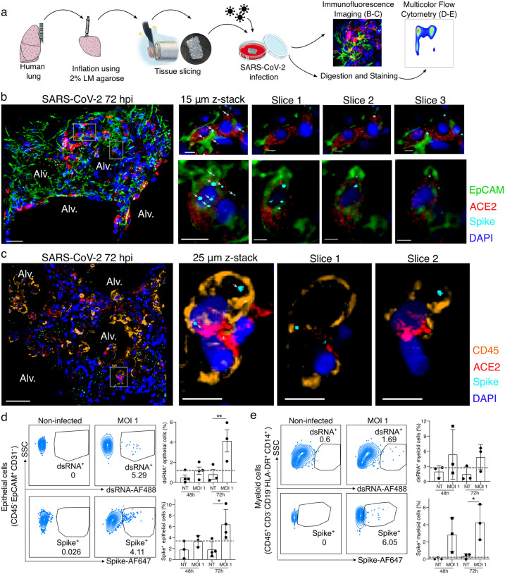
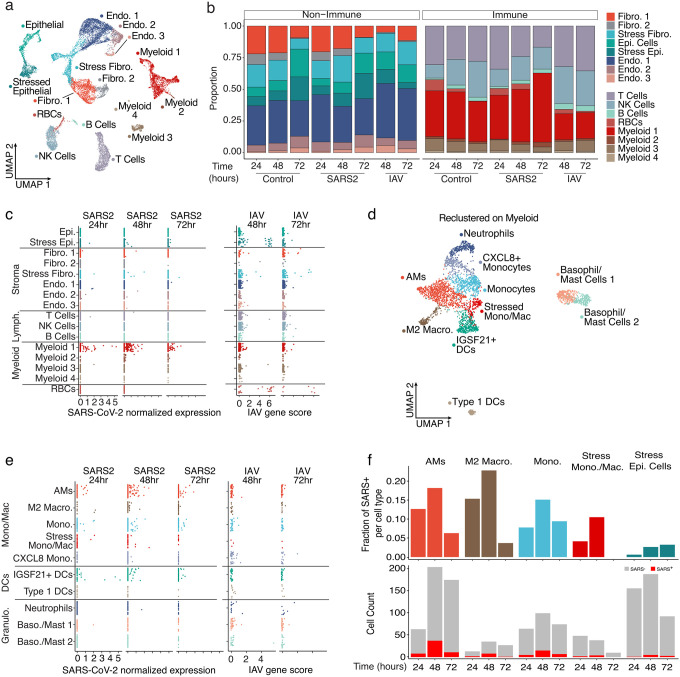
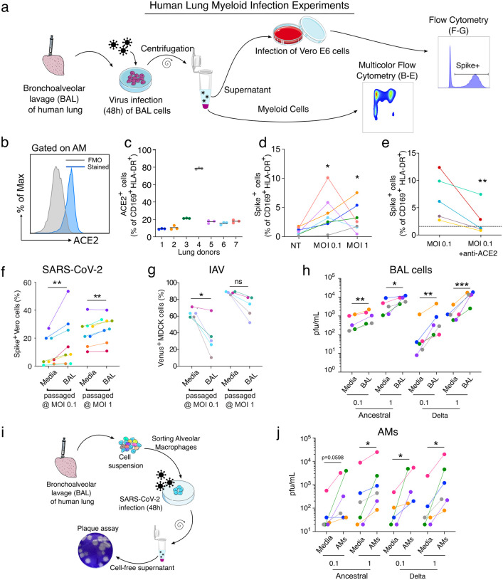
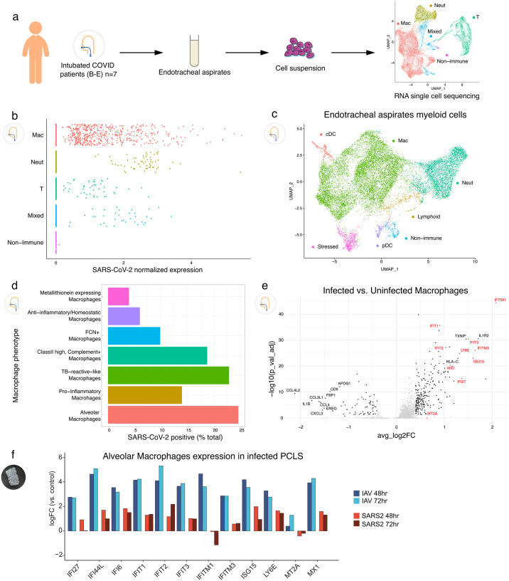
In the severe acute respiratory syndrome coronavirus 2 (SARS-CoV-2) pandemic, considerable focus has been placed on a model of viral entry into host epithelial populations, with a separate focus upon the responding immune system dysfunction that exacerbates or causes disease. We developed a precision-cut lung slice model to investigate very early host-viral pathogenesis and found that SARS-CoV-2 had a rapid and specific tropism for myeloid populations in the human lung. Infection of alveolar macrophages was partially dependent upon their expression of ACE2, and the infections were productive for amplifying virus, both findings which were in contrast with their neutralization of another pandemic virus, Influenza A virus (IAV). Compared to IAV, SARS-CoV-2 was extremely poor at inducing interferon-stimulated genes in infected myeloid cells, providing a window of opportunity for modest titers to amplify within these cells. Endotracheal aspirate samples from humans with the acute respiratory distress syndrome (ARDS) from COVID-19 confirmed the lung slice findings, revealing a persistent myeloid depot. In the early phase of SARS-CoV-2 infection, myeloid cells may provide a safe harbor for the virus with minimal immune stimulatory cues being generated, resulting in effective viral colonization and quenching of the immune system.


Conflict of interest statement

Competing interests. The authors declare no competing interests.

Figures

Figure 1.
Figure 1.. SARS-CoV-2 infects both epithelial and immune cells in human PCLSs.
(a) Schematic diagram of the experimental design. Human lung lobes were inflated with 2% low-melting point agarose and sectioned into 300 μm precision cut lung slices (PCLS), which were cultured in 24-well plates and infected with SARS-CoV-2 for 48 or 72h. After incubation, PCLS were either fixed and stained for confocal imaging (b, c) or dissociated and stained for flow cytometry (d, e). (b, c) PCLS were infected with SARS-CoV-2 for 72h at MOI 0.1 or 1 and used for confocal imaging. Alveolar spaces (Alv.) are indicated in the large image (scale bar = 50 μm). Zoom area is marked by the white rectangle. For each zoomed area, 15 or 25 μm z-stacks appears on the side with single x-y sections (scale bar = 10 μm). (b) PCLS were stained for DAPI (dark blue), EpCAM (green), ACE2 (red) and spike (light blue), (c) PCLS were stained for DAPI (dark blue), CD45 (orange), ACE2 (red) and spike (light blue), (d, e) PCLS were infected at MOI 1 for 48 and 72h. PCLS were dissociated and cell suspension was stained for flow cytometry analysis (n=3–4). Infection was assessed by intracellular spike and dsRNA staining in (d) epithelial and (e) myeloid cells. Grey dashed lines indicate detection limit of assays. Data are mean ± SEM. Each dot represents the average percentage of dsRNA+ or spike+ myeloid cells of 2 to 3 individual lung slices from one donor. *p<0.05, **p<0.01.
Figure 2.
Figure 2.. SARS-CoV-2 displays tropism for myeloid cells compared to IAV.
(a) A Uniform Manifold Approximation and Projection (UMAP) visualization of cells from control, SARS-CoV-2, and IAV-infected PCLS collected at distinct times, (b) Relative quantification of cell types from the different experimental conditions, stratified by timepoint. (c) Scatterplots describing the library-normalized SARS-CoV-2 expression across various cell types in SARS-CoV-2-infected PCLS (left) or IAV gene score in IAV-infected PCLS (right), (d) A UMAP of finely annotated myeloid cell types in the dataset, (e) Distribution of infected myeloid cells similar to (c). (f) The fraction (top) and numbers of SARS-CoV-2-positive cells (bottom) at different timepoints.
Figure 3.
Figure 3.. SARS-CoV-2 infection of alveolar macrophages is ACE2-dependent and amplifies viral titer,
(a) Bronchoalveolar lavage (BAL) of human lungs yields cells that were infected with SARS-CoV-2 for 48h. Infected cells were analyzed by multicolor flow cytometry. Supernatant of infected BAL cells was used to infect Vero E6 cells. 24h after infection, infected cells were quantified by flow cytometry, (b) ACE2 protein expression was assessed on alveolar macrophages (AMs: CD169+ HLADR+ cells) by flow cytometry, (c) Proportion of ACE2+ AMs was measured (n=7). (d) AM infection was measured by flow cytometry using spike staining (each color represents a human lung donor, n=7). (e) ACE2 blocking antibody was added to BAL cells before infection (MOI 0.1). Cells were analyzed by flow cytometry (n=5). (f) BAL cells were infected with SARS-CoV-2 (BAL passaged). As a control, SARS-CoV-2 was incubated in culture media alone (Media). Cell-free supernatant was used to infect Vero E6 cells. At 24h after infection, Vero E6 cells were stained for intracellular spike expression and analyzed using flow cytometry (n=8). (g) Similarly, IAV-Venus was used to infect BAL cells or incubated with media. Cell-free supernatant was used to infect MDCK cells. At 24h after infection, Venus expressing MDCKs percentage was measured by flow cytometry (n=5). (h) Plaque assay was used to further assess viral titer in supernatant of infected BAL cells (n=5–6). (i) AMs were sorted from BAL samples, (j) Following 48h of SARS-CoV-2 infection (Ancestral, delta), viral titer was determined by plaque assay (n=5–6). ns=not significant, *p<0.05, **p<0.01, ***p<0.001
Figure 4.
Figure 4.. Tropism of SARS-CoV-2 for myeloid cells in endotracheal aspirates from COVID-19 subjects with ARDS,
(a) Endotracheal aspirates were collected from SARS-CoV-2 infected subjects with ARDS and subjected to scRNA-seq. UMAP at far right shows landmark populations, (b) Distribution of per-cell normalized SARS-CoV-2 expression in landmark cell types, (c) UMAP projection of myeloid subtypes in endotracheal aspirates, (d) Fraction of SARS-CoV-2 positive cells per myeloid cell type (e) A volcano plot of SARS-Cov-2 positive AMs vs uninfected AMs. Interferon stimulated genes (ISGs) are highlighted in red. (f) Log2 Fold Change of select ISGs in the PCLS experiment in IAV- and SARS-CoV-2-infected cells at 48 and 72h vs control.

References

    1. Zhou P. et al. A pneumonia outbreak associated with a new coronavirus of probable bat origin. Nature 579, 270–273, doi:10.1038/s41586-020-2012-7 (2020). - DOI - PMC - PubMed
    1. Combes A. J. et al. Global absence and targeting of protective immune states in severe COVID-19. Nature 591, 124–130, doi:10.1038/s41586-021-03234-7 (2021). - DOI - PMC - PubMed
    1. Mathew D. et al. Deep immune profiling of COVID-19 patients reveals distinct immunotypes with therapeutic implications. Science 369, doi:10.1126/science.abc8511 (2020). - DOI - PMC - PubMed
    1. Rodda L. B. et al. Functional SARS-CoV-2-Specific Immune Memory Persists after Mild COVID-19. Cell 184, 169–183 e117, doi:10.1016/j.cell.2020.11.029 (2021). - DOI - PMC - PubMed
    1. Thornton E. E., Krummel M. F. & Looney M. R. Live imaging of the lung. Curr Protoc Cytom Chapter 12, 12 28 11–12 28 12, doi:10.1002/0471142956.cy1228s60 (2012). - DOI - PubMed

Publication types