. 2022 Feb 16:9:rbac008.
doi: 10.1093/rb/rbac008. eCollection 2022.

Fabrication and evaluation of a BMP-2/dexamethasone co-loaded gelatin sponge scaffold for rapid bone regeneration

Affiliations

Fabrication and evaluation of a BMP-2/dexamethasone co-loaded gelatin sponge scaffold for rapid bone regeneration

Qi Gan et al. Regen Biomater. .

Abstract

Improving the osteogenic activity of BMP-2 in vivo has significant clinical application value. In this research, we use a clinical gelatin sponge scaffold loaded with BMP-2 and dexamethasone (Dex) to evaluate the osteogenic activity of dual drugs via ectopic osteogenesis in vivo. We also investigate the mechanism of osteogenesis induced by BMP-2 and Dex with C2C12, a multipotent muscle-derived progenitor cell. The results show that the gelatin scaffold with Dex and BMP-2 can significantly accelerate osteogenesis in vivo. It is indicated that compared with the BMP-2 or Dex alone, 100 nM of Dex can dramatically enhance the BMP-2-induced alkaline phosphatase activity (ALP), ALP mRNA expression and mineralization. Further studies show that 100 nM of Dex can maintain the secondary structure of BMP-2 and facilitate recognition of BMP-2 with its receptors on the surface of C2C12 cells. We also find that in C2C12, Dex has no obvious effect on the BMP-2-induced Smad1/5/8 protein expression and the STAT3-dependent pathway, but Runx2-dependent pathway is involved in the Dex-stimulated osteoblast differentiation of BMP-2 both in vitro and in vivo. Based on these results, a potential mechanism model about the synergistic osteoinductive effect of Dex and BMP-2 in C2C12 cells via Runx2 activation is proposed. This may provide a theoretical basis for the pre-clinical application of Dex and BMP-2 for bone regeneration.

Keywords: BMP-2; Runx2; bone regeneration; dexamethasone; pre-clinical.


Figures

None
Graphical abstract
Figure 1.
Figure 1.
Effect of Dex on BMP-2-induced ectopic osteogenesis in vivo. (a) Gelatin sponge scaffold with 15 μg of BMP-2 and 0, 4, 20, 100 μg Dex was implanted into thigh muscle pouches of mice to stimulate ectopic osteogenesis. The digital pictures (b) and micro-CT images (c) of new ectopic bone after 2 weeks of surgery were displayed (n = 6). Use GE software-Microview ABA 2.1.2 to quantitatively analyze the new bone volume (d) and tissue mineral density (e) from the micro-CT image. The data presented are the average of three independent experiments. (f) Western blot analysis of Runx2 and β-actin protein abundances in ectopic bone formation. (g) Integrated in vivo Runx2 optical data analysis by ImageJ 1.42. * indicates that group with BMP-2 and 20 μg Dex contained higher bone volume than groups without Dex. # indicates that the tissue mineral density of groups with Dex and BMP-2 all higher than groups without Dex. & indicates that the Runx2 protein levels in vivo with Dex and BMP-2 higher than BMP-2 group. @ indicates that the Runx2 protein expression of the group with BMP-2 and 20 μg Dex is significantly higher than the other groups. *P, #P, &P, @P <0.05
Figure 2.
Figure 2.
Dex enhanced the biological activity of BMP-2. (a) ALP was measured after cells were cultured with 0.4 μg/ml of BMP-2 and 0, 5, 10, 20, 100, 200 or 500 nM of Dex for 3 days. The data were obtained from the color reaction of p-nitrophenylphosphate. (b) The staining of ALP activity was using fast blue BB and naphthol as-BI phosphate salt. (c) ALP activity of C2C12 cells for different incubation times. Cells incubated with BMP-2 (0.4 μg/ml), Dex (100 nM) or a combination of both for different incubation times. (d) The ALP activity was induced by adding inducers at different time periods. For the first 2 days of C2C12 cell culture, used BMP-2 (0.4 μg/ml), Dex (100 nM) or a mixture of two inducers as the medium, and removed the inducer after 48 h of culture. Then, we cultured the cells with the designated inducers from Day 3 to 4. ALP activity was measured at Day 4 (n = 4). (e) Effects of BMP-2 and Dex on C2C12 cells mineralization. The culture medium of C2C12 cells were BMP-2 (0.4 μg/ml), Dex (100 nM) or a mixture of two inducers. The staining of cell mineralization was detected using 1% Alizarin Red S and checked at 400× magnification. *P <0.05. **P <0.001
Figure 3.
Figure 3.
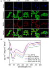
(a) Effect of Dex on the binding of BMP-2 to C2C12 cell membranes. The cells were incubated with 0.4 μg/ml BMP-2 and different concentrations of Dex for 6 h. FITC-labeled phalloidin (green) was used to stain the cell cytoskeleton and DAPI (blue) was used to stain the nuclei. BMP-2 on the cell surface was detected with alexa fluor 647-labeled anti-mouse IgG (red) and anti-BMP-2 antibodies. (b) Far-UV CD spectrum of BMP-2 at different concentrations of Dex. Secondary structure of BMP-2 was detected with related software
Figure 4.
Figure 4.
Effects of Dex on osteoblastic differentiation induced by BMP-2. (a) Effects of Dex on Smad1/5/8 phosphorylation signaling pathway induced by BMP-2. Western blotting for phosphorylated Smad1/5/8 was detected andβ-actin was set as the blank group. (b) Integrated p-Smads optical data analysis by ImageJ 1.42. (c) Western blot analysis of Runx2 and β-actin in C2C12 cells in vitro. (d) Integrated Runx2 optical data analysis by ImageJ 1.42. (e) Detection of the expression of id-1 affected by Dex and BMP-2. The mRNA expression of id-1 was measured with RT-PCR (n = 4). (f) ALP activity of C2C12 cells cultured with Dex and BMP-2 and the JAK/STAT inhibitor AG490. The concentration of BMP-2 in all experiments is 0.4 μg/ml and Dex is 100 nM. **P <0.001, *P <0.05
Figure 5.
Figure 5.
The expression of osteoblast markers and osteogenic transcription factors affected by BMP-2 and Dex, (a) ALP, (b) OC, (c) collagen I and (d) Runx2, in C2C12 cells. Cells were incubated with BMP-2 (0.4 μg/ml), Dex (100 nM) or a mixture of two inducers for 1, 3 and 5 days and characterized with RT-PCR (n = 4). Control indicates the cells cultured in a medium without BMP-2 and Dex. *P <0.05, ** P <0.001
Figure 6.
Figure 6.
Cartoon model showing the regulation of Dex on the binding of BMP-2 to its receptor and BMP-2-induced osteoblastic differentiation via Runx2 activation. Dex at low concentration is beneficial to the ability of BMP-2 to bind to cell membrane surface receptors and thus up-regulated the downstream signaling pathway. Dex has no obvious effect on phospho-Smad1/5/8 protein and id-1, but it can up-regulate Runx2 expression and then promote osteoblastic differentiation and inhibit myoblast differentiation in C2C12 cell model

Similar articles

Cited by

References

    1. Bolland B, Tilley S, New AMR, Dunlop DG, Oreffo ROC. Adult mesenchymal stem cells and impaction grafting: a new clinical paradigm shift. Expert Rev Med Devices 2007;4:393–404. - PubMed
    1. Freeman FE, Pitacco P, van Dommelen LHA, Nulty J, Browe DC, Shin J-Y, Alsberg E, Kelly DJ. 3D bioprinting spatiotemporally defined patterns of growth factors to tightly control tissue regeneration. Sci Adv 2020;6:5093. - PMC - PubMed
    1. Herberg S, McDermott AM, Dang PN, Alt DS, Tang R, Dawahare JH, Varghai D, Shin JY, McMillan A, Dikina AD, He F, Lee YB, Cheng Y, Umemori K, Wong PC, Park H, Boerckel JD, Alsberg E. Combinatorial morphogenetic and mechanical cues to mimic bone development for defect repair. Sci Adv 2019;5:2476. - PMC - PubMed
    1. McDermott AM, Herberg S, Mason DE, Collins JM, Pearson HB, Dawahare JH, Tang R, Patwa AN, Grinstaff MW, Kelly DJ, Alsberg E, Boerckel JD. Recapitulating bone development through engineered mesenchymal condensations and mechanical cues for tissue regeneration. Sci Transl Med 2019;11:7756. - PMC - PubMed
    1. Chen D, Zhao M, Mundy GR.. Bone morphogenetic proteins. Growth Factors 2004;22:233–41. - PubMed