Review
. 2022 May 3:10:883491.
doi: 10.3389/fcell.2022.883491. eCollection 2022.

Image-Based Analysis Revealing the Molecular Mechanism of Peroxisome Dynamics in Plants

Affiliations
Review

Image-Based Analysis Revealing the Molecular Mechanism of Peroxisome Dynamics in Plants

Shino Goto-Yamada et al. Front Cell Dev Biol. .

Abstract

Peroxisomes are present in eukaryotic cells and have essential roles in various biological processes. Plant peroxisomes proliferate by de novo biosynthesis or division of pre-existing peroxisomes, degrade, or replace metabolic enzymes, in response to developmental stages, environmental changes, or external stimuli. Defects of peroxisome functions and biogenesis alter a variety of biological processes and cause aberrant plant growth. Traditionally, peroxisomal function-based screening has been employed to isolate Arabidopsis thaliana mutants that are defective in peroxisomal metabolism, such as lipid degradation and photorespiration. These analyses have revealed that the number, subcellular localization, and activity of peroxisomes are closely related to their efficient function, and the molecular mechanisms underlying peroxisome dynamics including organelle biogenesis, protein transport, and organelle interactions must be understood. Various approaches have been adopted to identify factors involved in peroxisome dynamics. With the development of imaging techniques and fluorescent proteins, peroxisome research has been accelerated. Image-based analyses provide intriguing results concerning the movement, morphology, and number of peroxisomes that were hard to obtain by other approaches. This review addresses image-based analysis of peroxisome dynamics in plants, especially A. thaliana and Marchantia polymorpha.

Keywords: Arabidopsis thaliana; Marchantia polymorpha; apem mutant; imaging; peroxisome; peup mutant.


Conflict of interest statement

The authors declare that the research was conducted in the absence of any commercial or financial relationships that could be construed as a potential conflict of interest.

Figures

FIGURE 1
FIGURE 1
Detection of peroxisomes in leaf cells. Fluorescence microscopic analysis of GFP (A) and electron microscopic analysis (B) were performed of transgenic A. thaliana (GFP-PTS1) expressing the fusion gene of GFP with PTS1 under the regulation of the constitutive promoter. (A) A lot of peroxisomes were visualized as spherical structures (Jedd and Chua, 2002; Mano et al., 2002; Mathur et al., 2002). Some representative peroxisomes are indicated by arrows. Bar, 20 µm. (B) Transmission electron microscopic observation of GFP-PTS1 plants (Mano et al., 2002). P, peroxisome; Mt, mitochondrion; Ch, chloroplast; V, vacuole. Bar, 1 µm.
FIGURE 2
FIGURE 2
GFP fluorescence in root tissue of the WT plant and apem mutants expressing the peroxisome marker GFP-PTS1 (Mano et al., 2004; Mano et al., 2006; Goto et al., 2011; Goto-Yamada et al., 2014a). Bars, 20 µm.
FIGURE 3
FIGURE 3
Peroxisome aggregation in peup mutants. Representative images of peroxisomes (green) and chloroplasts (magenta) in leaf mesophyll cells of the WT plant and peup mutants (Shibata et al., 2013; Goto-Yamada et al., 2019). Peroxisomes associate with chloroplasts in the WT plant, whereas peroxisomes partially form aggregates in peup mutants. Bars, 10 µm.
FIGURE 4
FIGURE 4
Schematic model of APEM protein functions in peroxisome proliferation, lipid metabolism, protein transport machinery, and quality control. (A) During peroxisome fission, DRP3A/APEM1 is recruited to the peroxisome division site together with DRP3B in a PEX11- and FIS1-dependent manner (Kao et al., 2018). DRP proteins are polymerized and constrict to divide peroxisomes. (B) PXN/APEM3 import NAD into the peroxisomal matrix and this is required for optimal fatty acid β-oxidation. (C) Peroxisomal matrix proteins are captured by the receptor PEX5 or PEX7. The PEX5-PEX7-cargo complex translocates to peroxisomes by binding to the docking complex consisting of PEX14 and PEX13/APEM2. The E2 ubiquitin ligase PEX4 and the E3 ligase PEX2/PEX10/PEX12 supposedly ubiquitinate PEX5 to export it from the peroxisomal membrane with/without the force generated by the APEM9/PEX15/PEX26-tethered AAA-ATPase PEX1-PEX6 complex. Experimental data support the interactions between PEX13 and PEX7 (Mano et al., 2006), PEX13 and PEX15/PEX26 (Li et al., 2014), and PEX7 and PEX12 (Singh et al., 2009). (D) Damaged and/or unwanted peroxisomal proteins are supposedly maintained or degraded by the chaperone/protease activity of LON2/APEM10 protein. Excess damaged proteins accumulate inside peroxisomes. Peroxisomes become oxidative upon catalase inactivation and aggregation, and these peroxisomes are targeted for pexophagy to be degraded in the vacuole (Shibata et al., 2013). ATG proteins, including ATG2/PEUP1, ATG18A/PEUP2, ATG7/PEUP4/PEUP22, and ATG5/PEUP17, are involved in this process.
FIGURE 5
FIGURE 5
Images of M. polymorpha and visualization of peroxisomes using Citrine- and mRFP1-fused proteins. Vegetative haploid life form (thallus) on agar plate (A) and vermiculite (B) (Shimamura, 2016). Female (C) and male (D) sexual organs from the haploid thallus of a female plant or a male plant, respectively (Chiyoda et al., 2008; Inoue et al., 2019). Bars, 1 cm. (E) Fluorescence in peroxisomes was observed in thallus epidermal cells expressing both pro35S:PTS2-Citrine and pro35S:mRFP1-PTS1 genes (Mano et al., 2018). Bars, 10 μm.
FIGURE 6
FIGURE 6
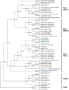
Phylogenetic relationships of PEX11 subfamilies. The numbers are the proportion of trees in which the associated sequences cluster together. Sequences of A. thaliana, M. polymorpha, and M. endlicherianum are colored as representatives from among angiosperms, bryophytes, and algae, respectively. The phylogenetic tree for PEX11 homologs was inferred using the Maximum Likelihood method and JTT matrix-based model (Jones et al., 1992) with MEGA11 (Stecher et al., 2020; Tamura et al., 2021). All positions with less than 95% site coverage were eliminated, i.e., fewer than 5% alignment gaps, missing data, and ambiguous bases were allowed at any position (partial deletion option). Orthologue sequences in plants were obtained from the datasets for C. braunii (Nishiyama et al., 2018), M. endlicherianum (Cheng et al., 2019), K. nitens (Hori et al., 2014), M. polymorpha (Montgomery et al., 2020), P. patens (Lang et al., 2018), A. agrestis (Li et al., 2020), S. moellendorffii (Banks et al., 2011), A. trichopoda (Amborella Genome Project, 2013), A. thaliana (Berardini et al., 2015), G. max (Schmutz et al., 2010), and T. aestivum (International Wheat Genome Sequencing Consortium, 2014). Other PEX11 species used in this analysis are P. pastoris (ANZ76138.1), S. cerevisiae (AJT75217.1), S. pombe (NP_595177.1), D. melanogaster (NP_611071.1), D. rerio (NP_001096590.1 and NP_001039319.1), H. sapiens (NP_003838.1 and NP_003837.1), and M. musculus (NP_035198.1 and NP_001155859.1).

Similar articles

Cited by

References

    1. Agne B., Meindl N. M., Niederhoff K., Einwächter H., Rehling P., Sickmann A. (2003). Pex8p: An Intraperoxisomal Organizer of the Peroxisomal Import Machinery. Mol. Cel 11 , 635–646.10.1016/s1097-2765(03)00062-5 - DOI - PubMed
    1. Amborella Genome Project (2013). The Amborella Genome and the Evolution of Flowering Plants. Science 342 (6165), 1241089. 10.1126/science.1241089 - DOI - PubMed
    1. Aung K., Hu J. (2012). Differential Roles of Arabidopsis Dynamin-Related Proteins DRP3A, DRP3B, and DRP5B in Organelle Division. J. Integr. Plant Biol. 54 (11), 921–931. 10.1111/j.1744-7909.2012.01174.x - DOI - PubMed
    1. Aung K., Hu J. (2009). The Arabidopsis Peroxisome Division Mutant Pdd2 Is Defective in the DYNAMIN-RELATED PROTEIN3A (DRP3A) Gene. Plant Signal. Behav. 4 (6), 542–544. 10.4161/psb.4.6.8699 - DOI - PMC - PubMed
    1. Baillie A. L., Falz A. L., Müller-Schüssele S. J., Sparkes I. (2020). It Started with a Kiss: Monitoring Organelle Interactions and Identifying Membrane Contact Site Components in Plants. Front. Plant Sci. 11, 517. 10.3389/fpls.2020.00517 - DOI - PMC - PubMed

LinkOut - more resources