

Retrosplenial Cortex Effects Contextual Fear Formation Relying on Dysgranular Constituent in Rats
- PMID: 35592254
- PMCID: PMC9112855
- DOI: 10.3389/fnins.2022.886858
Retrosplenial Cortex Effects Contextual Fear Formation Relying on Dysgranular Constituent in Rats
Abstract
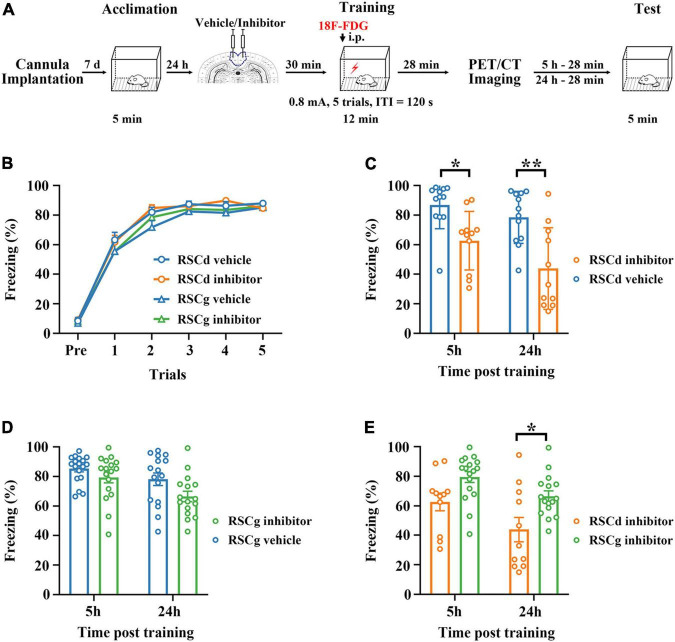
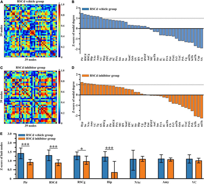
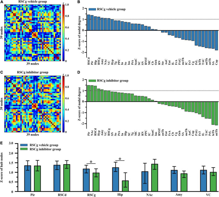
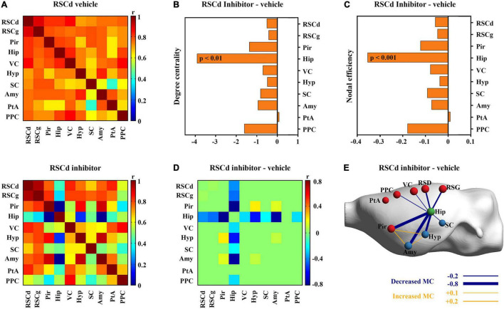
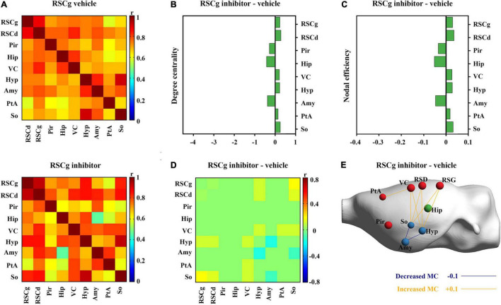
Animal contextual fear conditioning (CFC) models are the most-studied forms used to explore the neural substances of posttraumatic stress disorder (PTSD). In addition to the well-recognized hippocampal-amygdalar system, the retrosplenial cortex (RSC) is getting more and more attention due to substantial involvement in CFC, but with a poor understanding of the specific roles of its two major constituents-dysgranular (RSCd) and granular (RSCg). The current study sought to identify their roles and underlying brain network mechanisms during the encoding processing of the rat CFC model. Rats with pharmacologically inactivated RSCd, RSCg, and corresponding controls underwent contextual fear conditioning. [18F]-fluorodeoxyglucose positron emission tomography/computed tomography (18F-FDG PET/CT) scanning was performed for each animal. The 5-h and 24-h retrieval were followed to test the formation of contextual memory. Graph theoretic tools were used to identify the brain metabolic network involved in encoding phase, and changes of nodal (brain region) properties linked, respectively, to disturbed RSCd and RSCg were analyzed. Impaired retrieval occurred in disturbed RSCd animals, not in RSCg ones. The RSC, hippocampus (Hip), amygdala (Amy), piriform cortex (Pir), and visual cortex (VC) are hub nodes of the brain-wide network for contextual fear memory encoding in rats. Nodal degree and efficiency of hippocampus and its connectivity with amygdala, Pir, and VC were decreased in rats with disturbed RSCd, while not in those with suppressed RSCg. The RSC plays its role in contextual fear memory encoding mainly relying on its RSCd part, whose condition would influence the activity of the hippocampal-amygdalar system.
Keywords: brain metabolic network; contextual fear formation; graph theory; hippocampal-amygdalar system; retrosplenial cortex.
Copyright © 2022 Pan, Liu, Li, Zhang, Zhang, Zhao, Zhou, Nie, Zhu, Xu and Liu.
Conflict of interest statement
The authors declare that the research was conducted in the absence of any commercial or financial relationships that could be construed as a potential conflict of interest.
Figures
Similar articles
-
Fear-context association during memory retrieval requires input from granular to dysgranular retrosplenial cortex.Neurobiol Learn Mem. 2019 Sep;163:107036. doi: 10.1016/j.nlm.2019.107036. Epub 2019 Jun 12. Neurobiol Learn Mem. 2019. PMID: 31201928
-
Distinct Contribution of Granular and Agranular Subdivisions of the Retrosplenial Cortex to Remote Contextual Fear Memory Retrieval.J Neurosci. 2022 Feb 2;42(5):877-893. doi: 10.1523/JNEUROSCI.1303-21.2021. Epub 2021 Dec 7. J Neurosci. 2022. PMID: 34876468 Free PMC article.
-
Retrograde and anterograde contextual fear amnesia induced by selective elimination of layer IV-Va neurons in the granular retrosplenial cortex (A29).Neurobiol Learn Mem. 2020 May;171:107229. doi: 10.1016/j.nlm.2020.107229. Epub 2020 Apr 11. Neurobiol Learn Mem. 2020. PMID: 32289450
-
Role of retrosplenial cortex in processing stress-related context memories.Behav Neurosci. 2018 Oct;132(5):388-395. doi: 10.1037/bne0000223. Epub 2018 Jun 7. Behav Neurosci. 2018. PMID: 29878804 Free PMC article. Review.
-
Hippocampal network oscillations at the interplay between innate anxiety and learned fear.Psychopharmacology (Berl). 2019 Jan;236(1):321-338. doi: 10.1007/s00213-018-5109-z. Epub 2018 Nov 11. Psychopharmacology (Berl). 2019. PMID: 30417233 Review.
Cited by
-
Parvalbumin-expressing basal forebrain neurons mediate learning from negative experience.Nat Commun. 2024 Jun 7;15(1):4768. doi: 10.1038/s41467-024-48755-7. Nat Commun. 2024. PMID: 38849336 Free PMC article.
-
Medial amygdalar tau is associated with anxiety symptoms in preclinical Alzheimer's disease.bioRxiv [Preprint]. 2024 Jun 3:2024.06.03.597160. doi: 10.1101/2024.06.03.597160. bioRxiv. 2024. Update in: Biol Psychiatry Cogn Neurosci Neuroimaging. 2024 Dec;9(12):1301-1311. doi: 10.1016/j.bpsc.2024.07.012. PMID: 38895308 Free PMC article. Updated. Preprint.
-
Medial Amygdalar Tau Is Associated With Mood Symptoms in Preclinical Alzheimer's Disease.Biol Psychiatry Cogn Neurosci Neuroimaging. 2024 Dec;9(12):1301-1311. doi: 10.1016/j.bpsc.2024.07.012. Epub 2024 Jul 25. Biol Psychiatry Cogn Neurosci Neuroimaging. 2024. PMID: 39059466
-
The effects of amyloidosis and aging on glutamatergic and GABAergic synapses, and interneurons in the barrel cortex and non-neocortical brain regions.Front Neuroanat. 2025 Feb 12;19:1526962. doi: 10.3389/fnana.2025.1526962. eCollection 2025. Front Neuroanat. 2025. PMID: 40012738 Free PMC article.
-
Unique Transcriptomic Cell Types of the Granular Retrosplenial Cortex are Preserved Across Mice and Rats Despite Dramatic Changes in Key Marker Genes.bioRxiv [Preprint]. 2024 Sep 17:2024.09.17.613545. doi: 10.1101/2024.09.17.613545. bioRxiv. 2024. PMID: 39345493 Free PMC article. Preprint.
References
-
- Berger T. W., Weikart C. L., Bassett J. L., Orr W. B. (1986). Lesions of the retrosplenial cortex produce deficits in reversal-learning of the rabbit nictitating membrane response: implications for potential interactions between hippocampal and cerebellar brain systems. Behav. Neurosci. 100 802–809. 10.1037/0735-7044.100.6.802 - DOI - PubMed
LinkOut - more resources
Full Text Sources