

Conjunctival leiomyosarcoma
- PMID: 35592361
- PMCID: PMC9112115
- DOI: 10.1016/j.ajoc.2022.101580
Conjunctival leiomyosarcoma
Abstract
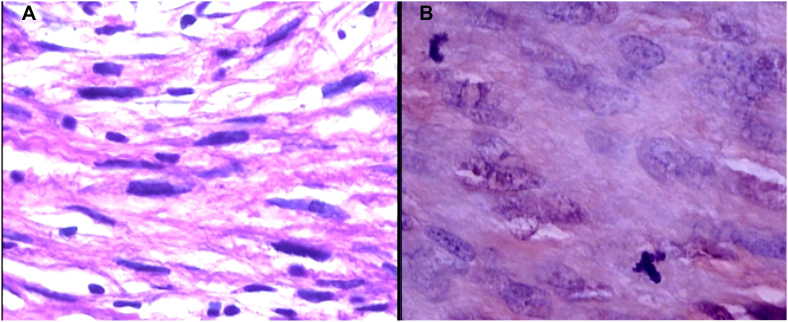
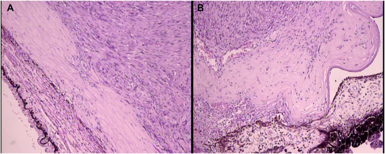
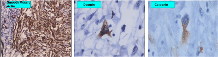
Purpose: Leiomyosarcoma (LMS) is a mesenchymal neoplasm with smooth muscle differentiation, being considered one of the most common soft tissue sarcomas. However, it rarely affects the eye, and when it does, it is usually located in the orbit, being extremely rare in the conjunctiva.
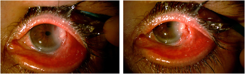
Observations: We report a case of a 45 years old male patient, with a recurrent rapid growing conjunctival mass on the temporal limbus of his left eye, which was excised, and the anatomopathological report was suggestive of a grade 1 leiomyosarcoma. Since the lesion was recurrent, we decided to perform an extended enucleation for treating this condition. Nevertheless, the patient is being followed up to 30 months, with systemic metastasis screening, showing no other lesions or recurrences.
Conclusions and importance: Conjunctival leiomyosarcoma is an extremely rare ocular tumor, which can be clinically indistinguishable from other conditions such as squamous cell carcinoma, so, biopsy is essential. Albeit there is no standard treatment, complete surgical removal with safety margins is mandatory.
Keywords: Conjunctiva; Eye; Leiomyosarcoma; Sarcoma; Soft tissue tumours; Tumor.
© 2022 The Author(s).
Figures
References
-
- Gotee J., Sioda N., Aurit S., Curtin C., Silberstein P. Important prognostic factors in leiomyosarcoma survival: a National Cancer Database (NCDB) analysis. Clin Transl Oncol. 2020;22(6):860–869. - PubMed
-
- Wolff-Rouendaal D. Xeroderma pigmentosum with ophthalmological symptoms. Ophthalmologica. 1976;173(3-4):290–291. - PubMed
-
- White V.A., Damji K.F., Richards J.S., Rootman J. Leiomyosarcoma of the conjunctiva. Ophthalmol. 1991;98(10):1560–1564. - PubMed
Publication types
LinkOut - more resources
Full Text Sources
