Intraosseous Lipoma of the Calvaria in the Early Stage Resembling Normal Fatty Marrow
- PMID: 35592430
- PMCID: PMC9113857
- DOI: 10.1055/s-0042-1747972
Intraosseous Lipoma of the Calvaria in the Early Stage Resembling Normal Fatty Marrow
Abstract
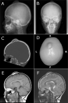
Intraosseous lipoma (IOL) is a benign bone tumor that usually arises from the lower limb and rarely arises from the skull. Radiological diagnosis of a typical case is not problematic due to its characteristic calcification and marginal sclerosis. Here, we report a case of calvarial IOL in the early stage lacking conventional radiopathological features. The patient is a 7-year-old girl who presented with a slow-growing protuberance on the vertex of the head. Computed tomography displayed a low-density mass without calcification that was continuous with the surrounding diploe. The mass was resected piece by piece for diagnostic and cosmetic reasons. Histologically, the specimen consisted of bony trabeculae and intertrabecular adipose tissue, which resembled normal fatty marrow. However, adipose tissue was considered neoplastic since it lacked hematopoietic elements. The final diagnosis of IOL was made by radiopathological correlation. This case suggests that IOL should be included in the differential diagnosis of diploic expansion, even if calcification is absent. The histology of an early-stage IOL resembles normal fatty marrow, but recognizing the absence of hematopoietic elements aids the diagnosis. Also, our literature review indicates that such cases are likely to be encountered in the calvaria than cranial base.
Keywords: calvaria; diploe; early; intraosseous; lipoma; skull.
The Author(s). This is an open access article published by Thieme under the terms of the Creative Commons Attribution-NonDerivative-NonCommercial License, permitting copying and reproduction so long as the original work is given appropriate credit. Contents may not be used for commercial purposes, or adapted, remixed, transformed or built upon. ( https://creativecommons.org/licenses/by-nc-nd/4.0/ ).
Conflict of interest statement
Conflict of Interest None declared.
Figures


References
-
- Chow L T, Lee K C. Intraosseous lipoma. A clinicopathologic study of nine cases. Am J Surg Pathol. 1992;16(04):401–410. - PubMed
-
- Unni K K, Inwards C Y, Research M FME. 6th edition. Wolters Kluwer Health/Lippincott Williams & Wilkins; Philadelphia, PA: 2009. Dahlin's Bone Tumors: General Aspects and Data on 10,165 Cases.
-
- Campbell R SD, Grainger A J, Mangham D C, Beggs I, Teh J, Davies A M. Intraosseous lipoma: report of 35 new cases and a review of the literature. Skeletal Radiol. 2003;32(04):209–222. - PubMed
-
- Propeck T, Bullard M A, Lin J, Doi K, Martel W. Radiologic-pathologic correlation of intraosseous lipomas. AJR Am J Roentgenol. 2000;175(03):673–678. - PubMed

