Case Reports
. 2022 May 12;11(5):20584601221102822.
doi: 10.1177/20584601221102822. eCollection 2022 May.

Pelvic schwannoma in an adult male

Affiliations
Case Reports

Pelvic schwannoma in an adult male

My Hanh Thi Dau et al. Acta Radiol Open. .

Abstract

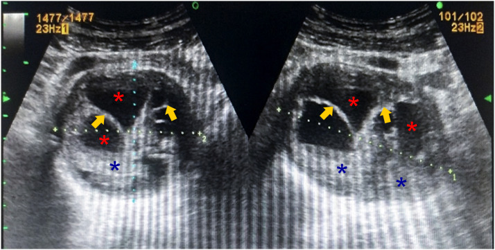
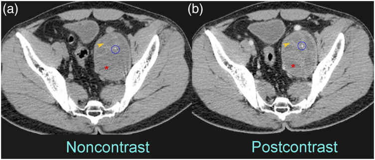
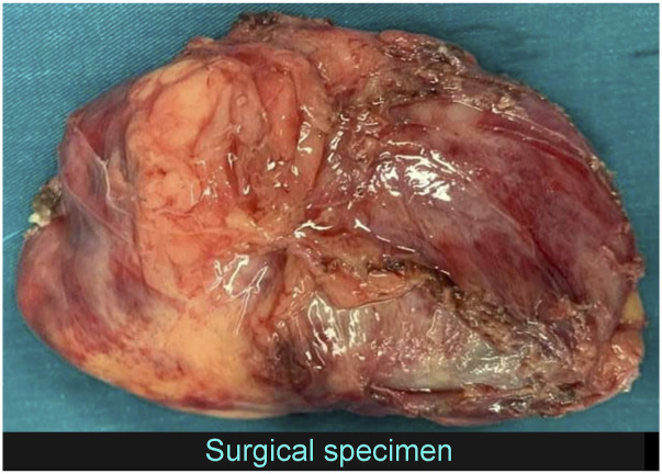
Schwannomas are benign nerve sheath tumors that are generally encapsulated and commonly detected in the head, neck, and mediastinal regions. Schwannomas localizing in the pelvis are extremely rare and tend to be asymptomatic initially due to slow growth rate. Schwannomas may be misdiagnosed as urologic or gynecologic tumors. Pelvic schwannomas are typically solitary, large, and well-circumscribed masses in the retroperitoneum or presacral areas. Other imaging characteristics are cystic degeneration, repeated hemorrhages, and calcifications. Calcification patterns can be punctate, speckled, curvilinear, or along the walls of the masses. We report a young man with a pelvic schwannoma with typical imaging features.

Keywords: Computed tomography; neurilemmoma; pelvic tumor; peripheral nerve sheath tumor; schwannoma.


Conflict of interest statement

Declaration of conflicting interests: The author(s) declared no potential conflicts of interest with respect to the research, authorship, and/or publication of this article.

Figures

Figure 1.
Figure 1.
Ultrasound images show a complex cystic lesion with internal septa (arrows), cystic (red asterisks), and solid (blue asterisks) components in the left hemipelvis.
Figure 2.
Figure 2.
CT images before (a) and after (b) contrast administration show a well-defined, heterogeneously enhancing mass with cystic areas (asterisks), hemorrhage (arrowheads), and small foci of calcification (circles) in the left lateral pelvis space.
Figure 3.
Figure 3.
MRI on T2-weighted image (a) and T1-weighted image (b) shows a well-defined, complex cystic lesion with hemorrhage (arrowheads), internal septa (arrows), and solid components (blue asterisks) in the left hemipelvis, and fat-saturated T1-weighted postcontrast (c) reveals heterogeneous enhancement of the internal septa and solid components.
Figure 4.
Figure 4.
Photograph of the gross specimen.

References

    1. Pak H, Haji Maghsoudi L. Pelvic Schwannoma: in light of a case report. Clin Case Rep 2019; 7: 2488–2490. - PMC - PubMed
    1. Nakashima T, Tsurumaru D, Nishimuta Y, et al. A case of pelvic schwannoma presenting prominent eggshell-like calcification. Case Rep Radiol 2013; 2013: 825078–825084. - PMC - PubMed
    1. Machairiotis N, Zarogoulidis P, Stylianaki A, et al. Pelvic schwannoma in the right parametrium. Int J Gen Med 2013; 6: 123–126. - PMC - PubMed
    1. Jindal T, Mukherjee S, Kamal MR, et al. Cystic schwannoma of the pelvis. Ann R Coll Surg Engl 2013; 95: e1–e2. - PMC - PubMed
    1. Hughes MJ, Thomas JM, Fisher C, et al. Imaging features of retroperitoneal and pelvic schwannomas. Clin Radiol 2005; 60: 886–893. - PubMed

Publication types

LinkOut - more resources